


Presurgical Planning Models
3D Printing
It has been 7 years since physicians at NewYork-Presbyterian Morgan Stanley Children’s Hospital became one of the first teams in the world to use 3D printing technology to produce a 3D replica of a child’s heart. A baby with borderline ventricle was spared 2 more surgeries as a result.
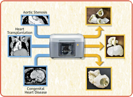
Since that first application of 3D printing, the Hospital’s pediatric heart surgeons and cardiac interventionalists have expanded their use of 3D pre-procedural planning models to guide surgery and interventional procedures in children born with complex heart lesions. These include transcatheter aortic valve replacement (TAVR), placement of VADs, and heart transplantation. Research by advanced imaging expert Dr. Kanwal M. Farooqi, Director of Cardiac 3D Printing and Imaging Director of the Initiative for Pediatric Cardiac Innovations, includes a multicenter study assessing the utility of 3D printed models for preoperative planning of VAD placement in patients with congenital heart disease and heart failure.
Three-dimensional printing technology is now increasingly being used as an advanced imaging technique to create unique patient-specific models for guidance prior to catheter-based or surgical interventions for patients with heart failure. A comprehensive review by Columbia faculty representing pediatric cardiology, radiology, pediatric cardiothoracic surgery, and mechanical engineering — published in JACC: Heart Failure, highlights the innovative applications of this technique to provide patient-specific models to improve patient care.