Elevating Precision and Safety in Cataract Surgery

Dr. Ashley Brissette and Dr. Kimberly C. Sippel
With the addition of femtosecond laser for cataract extraction and two innovative optical systems for measuring the refractive power of the eye during surgery itself, the Department of Ophthalmology at NewYork-Presbyterian/Weill Cornell Medical Center is able to offer patients visual results that have been documented to exceed national averages. Several new revolutionary devices assist in a) the precise performance of cataract surgery by femtosecond laser, and b) the intraoperative re-verification and precise positioning of the optimal intraocular lens for a given patient with the ability to correct for any surgically induced changes that may occur during the procedure. These and other remarkably powerful devices are housed in the three state-of-the-art ophthalmic operating suites in the NewYork-Presbyterian David H. Koch Center, acknowledged to be among the most advanced ophthalmic operating suites in the world.
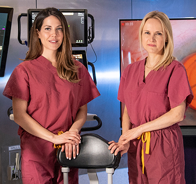
Corneal transplant surgeons Kimberly C. Sippel, MD and Ashley Brissette, MD, MSc share their perspectives on how these new technologies are transforming the practice of cataract surgery.
Precise, Safe, and Non-Invasive
Femtosecond laser-assisted cataract surgery is a non-invasive technique that replaces the least predictable and technically most demanding steps of conventional cataract procedures. A computer-guided laser that is linked to an optical imaging system performs the corneal incision, capsulotomy, and lens fragmentation steps, bringing a new level of precision and accuracy to specific steps in cataract surgery traditionally performed with hand-held surgical instruments.
“Laser cataract surgery makes us better surgeons by greatly reducing human variability from the surgery. Using the femtosecond laser allows us to make a perfectly centered, precisely sized capsulotomy with unmatched reliability and safety.” — Dr. Kimberly C. Sippel
“The femtosecond laser enables us to give our patients the very best cataract surgery possible,” notes Dr. Sippel. “Laser cataract surgery makes us better surgeons by greatly reducing human variability from the surgery. An early step in cataract surgery – called capsulotomy – is to make a circular ‘manhole cover’ opening in the cataract, and this maneuver is among the most difficult portions of the procedure. Using the femtosecond laser allows us to make a perfectly centered, precisely sized capsulotomy with unmatched reliability and safety.”
Laser-assisted cataract surgery eliminates the need for blades by delivering computer-guided laser pulses that incise ocular tissues with precision in the microns. In addition to capsulotomy, the femtosecond laser can also make microincisions in the cornea to correct astigmatism, and can also be used to fragment the central nucleus of the cataract – the densest part of the lens that requires the most energy to remove by traditional methods – and make its extraction easier and less traumatic for the eye. “The laser can accomplish all of these steps in seconds while the surgeon monitors the process on the laser screen,” says Dr. Brissette. “After the surgeon initially enters the desired laser surgical plan for a given patient, the laser swiftly carries it out with the patient in total comfort.”
“Cataract surgery has had an impressive evolution,” adds Dr. Sippel. “It’s one of the only surgical procedures where there is almost no resemblance to how it was performed 30 years ago. With the development of laser instrumentation, we can make the incision and lens opening with a more reliable, repeatable, and precise incision. The laser breaks up the cataract using less energy than ultrasound, which is less traumatic for delicate ocular tissue, and why we are seeing faster visual recovery.”
“The femtosecond laser has minimized any potential complications that could occur because it enhances the precision of the surgery. Some studies show that there also might be improved time to healing in these patients with complex medical conditions and ocular diseases.” — Dr. Ashley Brissette
Dr. Brissette also emphasizes the higher level of safety afforded by femtosecond laser surgery. “Having access to these advanced ocular technologies can make the surgery safer for a certain subset of patients that have complex eye conditions who also need cataract or corneal surgery,” she says. “The technology helps to minimize complications that could arise, for example, in patients who have comorbid eye diseases such as Fuchs’ dystrophy and those who have had previous eye trauma that can make the cataract less stable. These might also include patients with complex diabetic retinal disease and other ocular conditions that could make visualization during the surgery and the cataract removal itself more difficult. The femtosecond laser has minimized any potential complications that could occur because it enhances the precision of the surgery. Some studies show that there also might be improved time to healing in these patients with complex medical conditions and ocular diseases.”
“This laser can also help us correct astigmatism in the eye,” adds Dr. Brissette. “We know that astigmatism can degrade the quality of the vision after surgery. And so, with this technology we have a way of not only removing the cataracts but also correcting astigmatism at the same time of surgery.”
Enhancing Accuracy in IOL Selection and Placement
“In addition to the femtosecond laser, the new surgical suites also provide for intraoperative aberrometry and digital access marking,” says Dr. Sippel. “These three technologies are elevating cataract surgery to another level.”
Weill Cornell ophthalmologists use an intraoperative aberrometer that provides real time, reliable data, and image guidance to more accurately select the intraocular lens (IOL) power and placement during cataract surgery. “This instrumentation allows us to measure the eye and then fine-tune the power selection of the intraocular lens to attain even better refractive results,” says Dr. Sippel. “As an example, we might select a +20 diopter lens based on formulas and on preoperative measurements, which is pretty accurate. But by using the aberrometer, we are able to obtain measurements while we’re in the operating room, and it may tell us that actually we should be using a +20.5 lens. This is a powerful tool that helps to improve postoperative visual outcomes, even with patients who have previously undergone laser vision correction or photorefractive keratectomy surgery.”
“Because intraoperative aberrometry enables us to take a measurement after the cataract is removed and while the operation is still ongoing, patient outcomes are measurably better,” adds Dr. Brissette. “The ultimate measure is patient satisfaction, and all patients want to see better, if not close to perfectly, without glasses after cataract surgery. With this technology, we can meet and more often exceed their expectations.”
During IOL implantation, surgeons take particular care to ensure that inaccurate preoperative measurement and intraoperative misalignment do not cause postoperative residual astigmatism. Because many patients may also require correction of additional astigmatism by the use of a custom astigmatic-correcting (“Toric”) lens, and because these lenses require placement in the precise clock hour orientation, intraoperative alignment of these lenses is critical. In the past, surgeons used a preoperative pen mark, applied to the patient in the holding area, to note the orientation desired; during insertion, the lens was rotated to match this mark. Now, with intraoperative optical verification, the visual results after cataract surgery with astigmatic-correcting lenses are greatly improved.
“Computer alignment, which is coupled to the aberrometry technology, allows us to eliminate human error,” says Dr. Brissette. “In addition to optical verification, these new technologies allow the use of preoperative and intraoperative photographs of the iris to assist in guaranteeing perfect lens alignment within the eye.”
As a result of these advances and capabilities, visual results after cataract extraction by NewYork-Presbyterian Weill Cornell surgeons have been documented to exceed national averages.
A recent review of all cataract surgeries available in the Weill Cornell Epic medical record database documented postoperative visual acuity results of 20/40 or better in 97 percent of patients who did not have pre-existing vision limiting comorbidities, such as macular degeneration, compared to the nationally published average in which 94.6 percent of patients attain such an acuity.
Dr. Brissette believes that the ocular technologies will only continue to improve, providing even better outcomes for patients. “Having access to this technology has helped us improve our research abilities,” she says. “Because the laser helps in terms of the accuracy of determining the lens implant power at the time of surgery, we can track those outcomes more specifically for our patients and for our studies. What is wonderful about ophthalmology and medicine in general is a desire for constant innovation. At Weill Cornell we’re on the leading edge of that innovation because we embrace technology, always seeking to improve ourselves as surgeons in order to attain the best outcomes for our patients.”


