


The esophagus functions as a conduit for food after it has been chewed and swallowed. Barrett's esophagus is a precancerous condition that affects about 1 percent of adults in the United States and is more common in men than in women. It is believed to be a consequence of long-standing gastroesophageal reflux disease (GERD) — also known as acid reflux or heartburn.
Barrett's esophagus occurs when the normal lining (epithelium) of the esophagus is replaced by tissue that is similar to what lines the intestine. This abnormal tissue in the esophagus is known as "intestinal metaplasia." If left untreated, over time the intestinal metaplasia can turn into more severely abnormal tissue, termed "esophageal dysplasia."
At the Center for Advanced Digestive Care (CADC) at NewYork-Presbyterian/Weill Cornell Medical Center, a team of doctors specializing in esophageal disorders coordinates the care of every patient with Barrett's esophagus. Our gastroenterologists and interventional endoscopists are able to offer the latest diagnostic approaches and minimally invasive treatments. Patients who require surgery benefit from the experienced Upper GI and thoracic surgeons at NewYork-Presbyterian/Weill Cornell.
All team members work together to provide seamless care to improve the health and quality of life of people with Barrett's esophagus.
Symptoms of Barrett's Esophagus
Barrett's esophagus itself does not cause symptoms. However, many individuals with Barrett's esophagus may have symptoms such as heartburn, indigestion, or a sour taste in the mouth — symptoms associated with GERD.
Barrett's Esophagus Diagnosis
Endoscopy
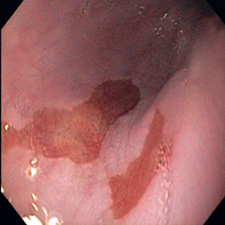
Barrett's esophagus is diagnosed by a gastroenterologist who performs an upper endoscopic examination of the esophagus — inspection of the inside of the esophagus using a flexible tube with a camera at its tip. The gastroenterologist is able to see abnormal areas of tissue which indicate Barrett's esophagus.
Tissue biopsies can then be taken of the area and are sent to a pathologist to review. The pathologist confirms the presence or absence of intestinal metaplasia and determines the degree of dysplasia (low, intermediate, or high grade).
Endoscopic Ultrasound
Endoscopic ultrasound is sometimes employed to assess Barrett's esophagus. This test is used to take a detailed look at the different layers of the esophagus. It is accomplished via an endoscope, just like routine upper endoscopy. The difference is the presence of an ultrasound probe at the tip of the scope.
Endoscopic ultrasound is performed in patients with "nodular" Barrett's esophagus. The term "nodular" refers to a raised area of tissue located near or within the Barrett's esophageal tissue. These raised or nodular areas are often a concern, because they may contain higher levels of dysplasia and possibly even cancer. The ultrasound can tell the gastroenterologist how deep a nodule extends into the esophagus, and whether it can be safely removed. It also permits the gastroenterologist to look at lymph nodes in the area and to sample them if they appear abnormal.
Chromoendoscopy
During chromoendoscopy, the doctor applies a stain or dye to tissue in the esophagus to enhance its appearance and distinguish between normal and abnormal tissue. Chromoendoscopy can aid in the diagnosis of Barrett's esophagus.
Narrow Band Imaging (NBI)
With this endoscopic technique, doctors use a special system to capture high-resolution images of the inner surface of the esophagus without the use of dyes. NBI relies on the fact that light of different wavelengths penetrates tissue at different depths. The longer the wavelength, the deeper the tissue penetration. Blue light penetrates superficially, while red light penetrates more deeply. By using light of different wavelengths, doctors can see fine features of tissue in the esophagus.
Barrett's Esophagus Treatment
The specialists at the CADC are experienced in diagnosing and treating esophageal disorders. An interdisciplinary team of gastrointestinal specialists in endoscopy, radiology, and surgery works together to provide each patient with coordinated, advanced, and individualized care.
There are many available treatments for Barrett's esophagus. The goal of these therapies is to preemptively remove or destroy the abnormal lining of the esophagus before the cells turn into esophageal cancer. These modalities include the following:
Radiofrequency Ablation (RFA)
A relatively newer therapy for Barrett's esophagus is radiofrequency ablation, or RFA. This method uses thermal (heat) energy to destroy the abnormal cells lining the esophagus. Because the heat energy does not penetrate deeply, RFA is a safe method to treat Barrett's esophagus. In addition, in recent studies, RFA has been proven to be effective over the long-term for patients with Barrett's esophagus that contains dysplasia. After three years, most patients experience complete disappearance of their Barrett's esophagus following RFA therapy.
Cryotherapy
The CADC offers cryotherapy, a new endoscopic procedure being used to help treat Barrett's esophagus. Cryotherapy involves the use of a super-cooled liquid or gas to freeze abnormal (dysplastic) cells found within Barrett's esophagus. It may also be used to treat patients in whom radiofrequency ablation has failed. More research remains to be done on the long-term effectiveness of cryotherapy, but results so far are very encouraging.
Endoscopic Mucosal Resection
Endoscopic mucosal resection (EMR) is a technique used to remove raised (nodular) or depressed areas of Barrett's esophagus. These areas have the most potential to contain cancer cells and also higher grades of dysplasia. Removal is achieved by placing a small rubber band around the tissue after it has been suctioned into a cap at the end of the endoscope. Once the area of concern has been banded, a "snare" is inserted and closed around the tissue. Electrocautery (heat) is then applied through the metal snare to cut the tissue out of the esophagus. The area is then allowed to heal, and in a few weeks, treatment with radiofrequency ablation can begin.
For more about interventional endoscopic procedures, visit the Advanced Interventional Endoscopy page.
Barrett's Esophagus Surgery
In the case of large lesions or those deep within the esophagus, some patients with Barrett's esophagus may need surgery to remove the abnormal tissue. The upper gastrointestinal and thoracic surgeons at NYP/Weill Cornell are highly skilled in performing the full range of surgical techniques for the esophagus and use minimally invasive approaches whenever possible, returning patients to their normal activities sooner.
Contact us
Call for an Appointment
NewYork-Presbyterian
Center for Advanced Digestive Care