


Fractures lines can be difficult to visualize after acute elbow injury, particularly in children. Below are eight sequential steps to aid in the radiographic recognition of occult signs of injury.
Steps: Hourglass sign/figure of eighty Anterior fat pad evaluation Posterior fat pad evaluation Anterior Humeral line Radio-capitellar line Inspection of the radial head Distal humerus examination Olecranon and ulnar examination
Step 1: Hourglass sign/figure of eighty
Search for an adequate "hourglass sign", or "figure of eight" at the distal humerus. If absent the study is not a true lateral and interpretation of steps 2 through 4 is less reliable.
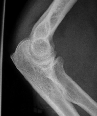
Here's an example of a true lateral; note the symmetric figure of eight/hourglass sign at the distal humerus; also notice the posterior fat pad? (see below)
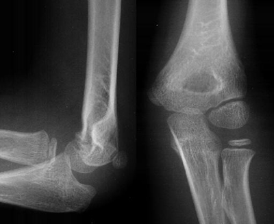
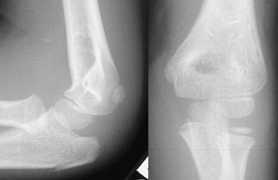
Here is an imperfect lateral radiograph accompanied by a normal AP radiograph; notice how the figure of eight/hourglass is asymmetric:
Step 2: Anterior fat pad evaluation
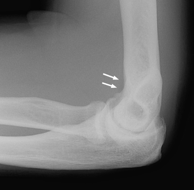
A visible anterior fat pad can be normal; it is a small radiolucent shadow adherent to the anterior aspect of the distal humerus:
An abnormal anterior fat pad is described as a "sail sign" because it is unusually prominent and bows outward to form a triangular shape. After trauma, blood can accumulate in the intraarticular space and push the fat pad anteriorly; a positive sail sign in the setting of trauma is a reliable indication of an intraarticular fracture – even if no fracture line can be identified. An atraumatic sail sign implies intraarticular fluid of an inflammatory nature.
Step 3: Posterior fat pad evaluation
Radiographic visualization of a posterior fat pad is never normal and always signifies fluid in the intraarticular space. Again, in the setting of trauma, this strongly implies fracture of an articular surface.
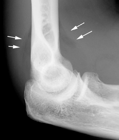
Here is a radiograph with both a sail and posterior fat pad sign:
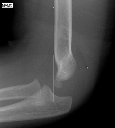
Step 4: Anterior Humeral line
This line should intersect the middle third of the capitellum on the lateral view. Fractures usually result in displacement of the capitellum posteriorly (versus anteriorly). If the film is not a true lateral, interpretation of the anterior humeral line becomes fallible.
This radiograph depicts a normal anterior humeral line:
This radiograph demonstrates abnormal alignment of the anterior humeral line strongly suspicious for fracture. (The anterior humeral line of a toddler/child must also intersect the middle third of an ossified capitellum; also note the posterior fat pad and sail sign.)
Step 5: Radio-capitellar line
This line is drawn through the middle of the radius posteriorly/rostrally and should bisect the capitellum on both the lateral and the AP elbow radiograph. Failure to align properly indicates a radial head dislocation that requires prompt reduction if neuro-vascular compromise is to be avoided.
Normal radio-capitellar lines
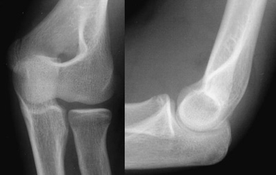
Notice that this is not an ideal lateral making interpretation of the anterior humeral line difficult; however the radius should bisect the capitellum on all views regardless of adequacy; also note the posterior fat pad.
An abnormal radio-capitellar line
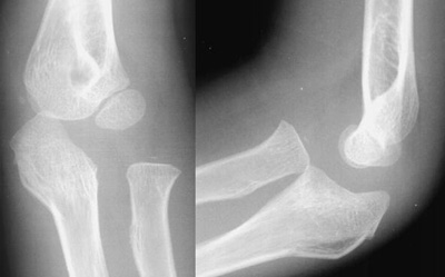
On both views the radius fails to bisect the capitellum indicating an obvious radial head dislocation. Also note the anterior and posterior fat pads, as well as the obvious olecranon deformity. A radial head dislocation with an olecranon fracture is called a Monteggia injury.
Step 6: Inspection of the radial head
Careful inspection is paramount since fracture lines are often not visible; look for subtle disruptions in the cortical contour. Examine the radiograph below:
Notice how the radius bisects the capitellum on this view; however there is a subtle cortical disruption/acute angulation at the superior aspect of the distal radius indicating fracture.
Step 7: Distal Humerus Examination
Signs of humeral fracture are also commonly subtle; breakage may only be evidenced by an abnormal anterior humeral line. Exam the radiograph below:

The anterior humeral line is perhaps slightly off as it seems to intersect the anterior third of the capitellum, while the radio-capitellar line is intact. There are prominent sail and posterior fat pad signs; and on careful inspection one sees the subtle cortical disruption along the posterior aspect of the distal humerus.
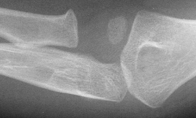
Step 8: Olecranon and ulnar examination
Look for obvious fracture lines and subtle disruptions in cortical contour.
This is not a good lateral radiograph so interpretation of the anterior humeral line is unreliable. There is no obvious anterior or posterior fat pad, and the radio-capitellar line is intact. The radial head and distal humerus appear fine, while there is an obvious proximal olecranon fracture.
Use this systematic approach to the elbow radiograph to avoid missing occult fractures of the elbow.