


3-D Printed Heart Models Give Surgeons Vital Information about a Heart's Anatomy
Pediatric heart surgeons and interventionalists at NewYork-Presbyterian are using advanced 3-D printed pre-surgical planning models to guide surgery in children born with complex heart lesions, with the goal of improving accuracy and outcomes.
Based on patient cardiac MRI and CT scans, the printer can produce an exact replica of the baby’s heart, allowing physicians to look at the inside of the heart in advance of surgery, providing them with more accurate information to precisely plan the surgery and allowing more time in the operating room to repair the child’s cardiac defects. Typically, surgeons must wait until they are in the operating room to see the extent of a heart’s congenital defect, and then execute the surgical procedure in a limited amount of time.
With the aid of a 3-D model, printed to scale, doctors are often able to repair all of a heart’s defects in a single procedure. Usually, babies born with complex defects require three or more life-threatening surgeries to make repairs.
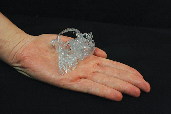
Thanks to this 3-D heart model, cardiac surgeons at NewYork-Presbyterian Congenital Heart Center could plan an infant’s operation before stepping into the OR.