Retinal Detachment: Improving Outcomes with Technique and Technology

Dr. Thanos Papakostas
The incidence of rhegmatogenous retinal detachment, the most common type of retinal detachment, has been reported to be between 6.3 and 18 per 100,000 each year. The age-specific incidence increases to a peak in both sexes in the 60- to 70-year age group.
“The estimated lifetime risk is 3 percent at 85 years of age. The risk increases in patients with high myopia, presence of lattice degeneration, trauma, or after cataract surgery. Retinal detachment is a major cause for universal vision loss if not treated quickly,” says Thanos Papakostas, MD, a retinal specialist in the Department of Ophthalmology at NewYork-Presbyterian/Weill Cornell Medical Center.
“The development of the small-gauge vitrectomy, wide-angle viewing systems, and the curved illuminated laser probe were key changes in the field.”
— Dr. Thanos Papakostas
According to Dr. Papakostas, the surgical techniques and technology currently available today have greatly improved outcomes in the treatment of retinal detachment. In many cases, speed in treatment is critical. “Macula-on retinal detachment is, essentially, an emergency,” says Dr. Papakostas. “We operate as soon as possible, usually on the same day or next day so that we can preserve the patient’s vision.”
While trauma is one cause for this form of retinal detachment, most happen spontaneously when the vitreous humor detaches from the retina. “As it detaches, it can cause a retinal break, the beginning of the detachment if left untreated,” says Dr. Papakostas. “The good news is that a retinal break can be addressed in the clinic with a minor laser procedure. But when it becomes a detachment, it is more involved. This is when you go to the OR.”
Dr. Papakostas performs one of several procedures to repair the detachment based on specific indications. Pneumatic retinopexy, performed in the office, involves injecting a gas bubble into the middle of the eye so that the bubble floats and presses against the retinal tear. The surgeon then seals the tear by applying cryotherapy or laser to the break.
“Other procedures include scleral buckling and vitrectomy; the indications for each is different depending on the unique characteristics of the retinal detachment,” says Dr. Papakostas. “Not all retinal detachments are the same. For example, a retinal detachment in a young patient is different than one in an elderly patient. We first consider the status of the lens in the eye. The patient can have a crystalline lens or an artificial lens, which happens with cataract surgery, or, in some cases, no lens.”

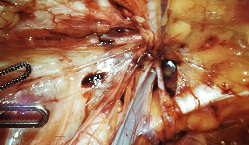
Retinal detachment associated with a giant retinal tear
The status of the vitreous also plays a role. “If the vitreous is attached, there is a crystalline lens, and if the patient is young, we tend to do a scleral buckle,” says Dr. Papakostas. In this procedure, a piece of silicone band is sewn onto the sclera at the site of the tear to push the sclera towards the retina, thus relieving the vitreous traction on the retina.
“The scleral buckle has been performed for years and has a very high success rate,” says Dr. Papakostas. “In general, however, if the patient is older with a detachment of the vitreous and has an artificial lens, we tend to do a vitrectomy.” In a vitrectomy, the vitreous humor is removed, the subretinal fluid is drained, and then laser is applied around the retinal breaks and a long-acting gas bubble is injected in the eye. “Vitrectomy allows us to treat complicated forms of retinal detachments that involve scar tissue formation and also retinal detachments after open-globe injuries.”
Technology Paves the Way
“Successful outcomes with vitrectomy are due, in large part, to advances in instrumentation technology,” says Dr. Papakostas. “The instrumentation used in the 1970s when this technique was developed was crude. Nowadays, we use very small instruments to enter the eye – 23-gauge, 25-gauge, and 27-gauge – that are atraumatic. They do a good job of removing as much gel as possible without causing any damage to the retina. The eye has minimal inflammation after the surgery as well.”
Today’s laser technologies also have enhanced surgical procedures. “The development of the curved illuminated laser probe was a key change in the field,” says Dr. Papakostas. “We can now treat the retina very safely without damaging the crystalline lens of the eye. Wide-angle viewing systems have also played a significant role in the development of vitrectomy surgery. These systems can provide wide-angle panoramic views of the retina that help the surgeon to identify all of the pathology, ensure a safe surgery, and decrease operating time.”
High-resolution, three-dimensional visualization is replacing direct optical viewing of the surgical field through a microscope. “The surgeon operates while wearing the 3-D fitted glasses and looking at a 4K high-definition monitor,” says Dr. Papakostas. “The monitor facilitates the involvement of the whole operating room team. Everyone can see exactly what the surgeon is seeing.
In addition, we can use significantly lower levels of illumination in the retina. We know from experimental data, as well as human data, that prolonged strong illumination on the surface of the retina can be toxic. And being able to sit comfortably through the surgery improves ergonomics for the surgeons, who are notorious for getting back and neck problems.”
A Closer Look at Complications
In addition to clinical practice, Dr. Papakostas is pursuing research focused on proliferative vitreoretinopathy (PVR). “Proliferative vitreoretinopathy is abnormal scarring inside the eye after retinal detachment repair and also the main reason for failure,” he says. “Currently, meticulous surgery is the standard treatment of PVR. Better understanding of the biology of this disease will lead to new drugs that will improve outcomes.”
Although scleral buckles can lead to high success rates in retinal detachment surgery, they can be associated with certain complications, such as change in the refractive error of the eye, diplopia, infection, and buckle extrusion, among others.
Most recently, Dr. Papakostas reviewed the postoperative complications of scleral buckling, the results of which were published in the November 2017 issue of Seminars in Ophthalmology. “Careful planning, appropriate patient selection, and good intraoperative technique can reduce the rate of these complications,” says Dr. Papakostas.
Reference Article
Papakostas TD, Vavvas D. Postoperative complications of scleral buckling. Seminars in Ophthalmology. 2017 Nov 29:1-5.
Related Publications

The Rarefied World of Ocular Oncology


