Pacesetters in Heart Rhythm Treatment

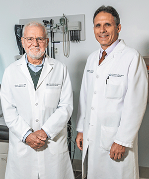
Dr. Hasan Garan and Dr. Angelo B. Biviano
Under the leadership of Hasan Garan, MD, the Cardiac Electrophysiology Department at NewYork-Presbyterian/ Columbia University Irving Medical Center is engaged in studies to better understand arrhythmias and their treatment. Cardiac electrophysiologist Angelo B. Biviano, MD, MPH, in collaboration with Columbia’s Adult Congenital Heart Center led by Marlon S. Rosenbaum, MD, has been pursuing studies involving patients with congenital heart disease and arrhythmias. “Patients with tetralogy of Fallot have a high prevalence of atrial tachyarrhythmia and often undergo ICD implantation at younger ages,” says Dr. Biviano. In a 20-year retrospective study of 40-plus patients with TOF, Columbia researchers found that a lower therapy cutoff rate for ventricular tachycardia was associated with increased risk of inappropriate shocks, the majority of which occurred in the presence of atrial arrhythmias. The authors advise that the risks of inappropriate shocks be considered when setting tachycardia therapy parameters in these patients.
In a study published in the April 5, 2019, issue of JACC Clinical Electrophysiology, Dr. Garan, Dr. Biviano, and their colleagues reported on the outcomes of 140 patients undergoing catheter ablation for atrial tachycardia. “We determined that catheter ablation in patients with adult congenital heart disease is effective in controlling arrhythmias,” notes Dr. Garan. “Specifically, acute procedural success was a predictor of freedom from recurrence, and the majority of patients achieved multiple arrhythmia-free years.”
Faculty also collaborated on a study looking at predictors and rates of recurrence of atrial arrhythmias following catheter ablation. Led by Matthew Lewis, MD, MPH, the study reviewed 124 patients treated with catheter ablations at Columbia over 10 years. The study did not find a significant difference in recurrence rates by gender, age, non-Fontan diagnosis, or need for transseptal puncture. However, BMI was a significant risk factor independent of Fontan status. These findings may help guide treatment decisions for persistent arrhythmias in the adult congenital heart disease population.

Dr. Elaine Y. Wan
Elaine Y. Wan, MD, a cardiac electrophysiologist, is developing in collaboration with Elisa E. Konofagou, PhD, who directs the Ultrasound Elasticity Imaging Laboratory in the Biomedical Engineering Department at Columbia University, electromechanical wave imaging (EWI), a novel, non-invasive technology to image cardiac tissue. “This modality uses ultrasound to image the origin of arrhythmias, including atrial tachycardia, atrial flutter, premature ventricular complexes, and accessory pathways in Wolff-Parkinson-White syndrome,” says Dr. Wan. “The ultrasound helps identify sources of the arrhythmias, which could be driven from either side of the atria, the right ventricle, or the left ventricle. Localization of arrhythmias is critical for treatment planning prior to cardiac ablation. The 12-lead electrocardiogram [ECG] can be limited in specificity and subject to interobserver variability. By using the ultrasound probe, we can take many more views of the heart, which we combine to generate a 3-D map of the electromechanical activation of heart rhythm.”
Dr. Wan and her colleagues first evaluated the procedure in 15 pediatric patients with 100 percent accuracy in predicting the location of the abnormal pathway. Preliminary studies presented at the late-breaking trials of the Heart Rhythm Society showed in a double-blinded study of 55 adult patients that the imaging technology was superior to the standard of care with ECG. EWI predicted 96 percent of arrhythmia locations as compared with 71 percent with ECG analyses. “We hope EWI may enable us to better predict procedural risks, reduce the amount of time it takes for the procedure, limit unnecessary radiation, and achieve a better outcome,” says Dr. Wan.


