


Comprehensive Management of Placenta Accreta
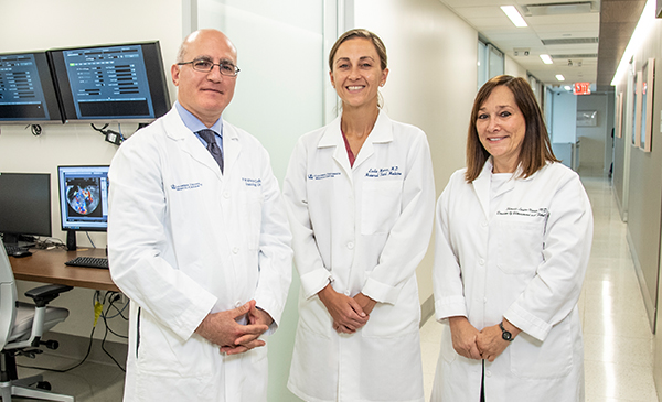
Dr. Fady Khoury-Collado, Dr. Leslie Moroz, and Dr. Sherelle Laifer-Narin
As the rate of Cesarean section deliveries increases in the United States, so, too, does the incidence of placenta accreta. “In 1950, the incidence was one in 30,000 deliveries; during the ‘80s and ‘90s it was about one in 730 deliveries. Today, it is about one in 500 deliveries,” says Leslie Moroz, MD, Director of the Mothers Center and Critical Care Obstetrics at NewYork-Presbyterian/Columbia University Irving Medical Center. Dr. Moroz and her colleagues Fady Khoury-Collado, MD, in the Department of Obstetrics and Gynecology, and Sherelle Laifer-Narin, MD, Department of Radiology, lead a comprehensive program to diagnose and manage these medically and surgically complex pregnancies.
Placenta accreta spectrum refers to the range of adherence of the placenta to the uterine muscle: placenta accreta is when the placenta attaches more firmly to the uterus and no longer separates spontaneously after the baby’s birth; placenta increta when it grows and becomes embedded in the uterine wall; and placenta percreta when it grows through the uterus and potentially to nearby organs. In any of these three situations, attempts to remove the placenta can lead to massive and life threatening hemorrhage. The risk for developing a placenta accreta spectrum increases with each Cesarean section or uterine surgery; other risk factors include placenta previa, advanced maternal age, multiparity, and uterine curettage.
“We spend a lot of time thinking about the best pathways of care for these very complicated patients,” says Dr. Moroz. “We receive referrals from our own healthcare system and patients are also referred by physicians at other institutions, especially smaller community hospitals that might not have the necessary resources.”
Importance of Imaging
“Our first step is to focus on the initial imaging with ultrasound and try to risk stratify those patients who have concerning findings. We also take a thorough medical history to determine the mother’s clinical risk factors,” continues Dr. Moroz, who notes that neither a patient’s clinical history or imaging studies are 100 percent predictive of the actual diagnosis. “We can identify increased risk based on either ultrasound or MRI or both, and certainly when they correlate, we become more concerned about the diagnosis. But one of the challenges of placenta accreta is that, to a certain extent, you don’t really know until the time of surgery exactly what the diagnosis will be.”
Sherelle Laifer-Narin, MD, an expert in both fetal and maternal diagnostic imaging, oversees ultrasound and fetal MRI in the Division of Abdominal Imaging. “If there is a question on ultrasound, an MRI can be performed to arrive at a more definitive diagnosis,” says Dr. Laifer-Narin, who generally performs imaging in the late second to early third trimester. “On an MRI, we can visualize the entire gestation on sequences of consecutive images. I can specifically localize where the placenta looks abnormal, and if any other organs, for example the bladder, are affected.”
If an abnormality is detected, Dr. Moroz begins to put together a tentative surgical plan to determine whether the patient will deliver in a labor and delivery operating room or in the Hospital’s main surgical operating room equipped with all of the necessary ancillary surgical services. “This would be based on a level of concern for the presence of placenta accreta spectrum and the anticipated surgical complexity,” says Dr. Moroz. “We also take additional precautions involving anesthesia. We anticipate the need for massive transfusion and place central lines as well as arterial lines, in the event of the need for close blood pressure monitoring, frequent lab draws, and the use of vasopressors.”
Sometimes, notes Dr. Moroz, conflicting results on imaging studies compel the team to wait until the time of surgery to make a definitive plan.
Comprehensive Planning for Complex Procedures
Gynecologic oncologist Fady Khoury-Collado, MD, specializes in surgeries for gynecologic cancers as well as the surgical management of patients with abnormal placentation. In collaboration with specialists in the Division of Maternal-Fetal Medicine, Dr. Khoury-Collado is a key member of the planning team. “I see the patients during their pregnancy to talk to them about my role in their care, which is doing the surgery to remove the uterus. After Dr. Moroz and her team deliver the baby, then my part starts,” says Dr. Khoury-Collado. “The main risk of placenta accreta is that an unplanned delivery and attempts at removing the placenta could lead to a catastrophic hemorrhage for the mother. The best way to manage this clinical condition is to know before the delivery that this condition is present and be prepared for it.”
As Dr. Khoury-Collado explains, the more the placenta invades the wall of the uterus, the greater the risk and difficulty in surgical management. “We try to prevent the bleeding by not delivering the placenta,” he says. “We deliver the baby only and then we remove the whole uterus with the placenta left in place. Despite that, there's still a high risk of bleeding that needs to be managed surgically. Our experience in managing these cases continues to evolve, and we have observed a concomitant steady improvement in patient outcomes.”
The multidisciplinary involvement of clinical services includes calling on urologists to perform a preoperative cystoscopy and bilateral ureteral stent placement in the event that the placenta has extended beyond the uterus into the space around the bladder. “We have that added level of protection to identify the anatomy in this area and to reduce the risk for urologic injury,” says Dr. Moroz. “In addition, our team of expert anesthesiologists focuses on the resuscitation of the patient during surgery, and their role is critical in replacing the blood loss in a timely fashion to maintain the patient stable throughout surgery and the postoperative period.”
The delivery is done in coordination with a mobile team from the Neonatal Intensive Care team since the majority of deliveries are performed in the late preterm period. “Thanks to the expertise of the obstetric anesthesia team, in many cases mothers are able to have their delivery under epidural anesthesia so they can be awake for the birth of their child,” adds Dr. Moroz. “Despite the complexity of the surgery, we try to preserve the joy of the delivery room.”
Addressing the Emotional Ramifications
Dr. Moroz and her Columbia colleagues also address the emotional impact on women as they face this life-threatening diagnosis and major surgery that presents such risks for mothers and their babies. “A diagnosis of placenta accreta fundamentally changes a woman’s thoughts about her pregnancy and pregnancy experience. There’s a lot of anxiety and it can be a huge challenge for the mothers. So, another aspect of prenatal care is making sure that they have the right kinds of support. We rely greatly on Dr. Catherine Monk, a clinical psychologist, and her mental health program within our department to provide services that include support groups and counseling to talk through some of the adjustment reaction that happens following a diagnosis like this. This also helps the mother come to terms with the ultimate delivery plan.”