NewYork-Presbyterian/Columbia physicians have developed a novel approach to the diagnosis and treatment of macular edema that harkens a new era of precision medicine for this debilitating condition. Macular edema, swelling of the retina, is a leading cause of vision loss in many intraocular and systemic diseases. Although optical coherence tomography (OCT) is one of the most accurate ways to diagnose the condition, the images offer little to help physicians assess the degree of retinal damage or predict a patient’s therapeutic response.
To address these unmet needs, Tongalp H. Tezel, MD, Director of the Vitreoretinal Service and Fellowship Program in the Department of Ophthalmology, and others at NewYork-Presbyterian/Columbia conducted a study using OTC to obtain cross-sectional images of the retina in various vision pathologies over time to characterize patterns of swelling in the retina. The findings led to groundbreaking insights about disease progression of macular edema and the development of a powerful algorithm that enables physicians to provide precision care that address patients’ individual needs.
“Several conditions, including diabetic retinopathy, age-related macular degeneration, and uveitis, can cause macular edema, the buildup of fluid in the retina,” explains Dr. Tezel. “The retina is sensitive tissue that captures light, processes it and sends the signal to the brain. When fluid accumulates in the retina, it scatters the light rays and defocuses the image on the retina, thereby impairing vision. The excess fluid can also compress the retina and cause cellular death, which can lead to or permanent vision loss.”
Assessing damage and therapeutic response prediction
Treatments for macular edema typically address the patient’s underlying condition (such as diabetes or inflammation) and the excess fluid leaking from blood vessels in the retina. These may include eye drops containing NSAIDs, intravitreal (IVI) injections containing anti-VEGF drugs or steroids, laser treatment, or surgery. A patient’s prognosis depends on many factors, including the duration and severity of the underlying condition, resulting retinal cell damage, and current vision status.
“Until now, there was no precise or internally standardized method to determine retinal hydration as an indicator of a patient’s retinal cell viability and whether they would benefit from a particular treatment,” explains Dr. Tezel. “Physicians tend to take a ‘one-size-fits-all’ approach to treating macular edema that is generalized to what is known about the disease, not to the individual patient.”
Until now, there was no precise or internally standardized method to determine retinal hydration as an indicator of a patient’s retinal cell viability and whether they would benefit from a particular treatment. Physicians tend to take a ‘one-size-fits-all’ approach to treating macular edema that is generalized to what is known about the disease, not to the individual patient.
— Dr. Tongalp Tezel
“The goal of this study was to develop a more precise method for diagnosing and treating macular edema,” he continues. “For every patient with this condition, we want to be able to answer important questions such as: How much fluid is inside the retina? What is the potential efficacy of different drugs to dehydrate the retina? What is the optimal time to treat, and how likely is the retina to recover sight?”
OCT is a non-invasive imaging technique that uses light waves to take cross-sectional images of the retina. Physicians use OCT to identify, quantify and monitor macular edema by measuring retinal thickness (RT). Although OCT can detect minute changes in RT, it cannot detect the beginnings of retinal cellular damage that leads to permanent visual loss, which can occur long before any change in RT vascular leakage occurs. Hence, timely and targeted intervention is necessary to prevent permanent visual loss.
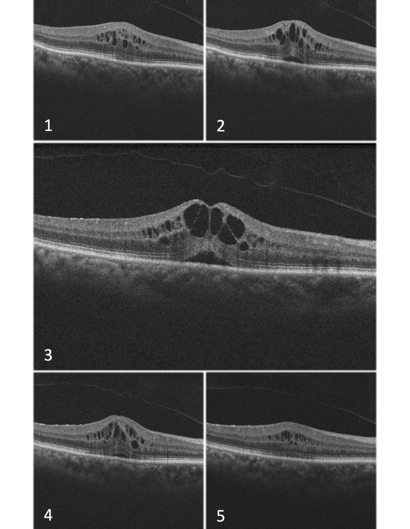
Harnessing the imaging power of OCT, Dr. Tezel and his study team theorized that characterizing tissue changes due to fluid accumulation in the retina as they occur through time could provide much-needed insights into the disease progression of macular edema. “Light reflection within the retina will diminish as it travels through pockets of fluid, and these areas will show as black on the OTC scan, or an optically empty area (OEA),” explains Dr. Tezel. “Thus, comparing the total black area within an OCT scan can give us an idea about the relative hydration of the sensory retina.”

Cystoid Macular Edema (CME) with optically empty areas (OEA), different sections of Optical Coherence Tomography (OCT) images
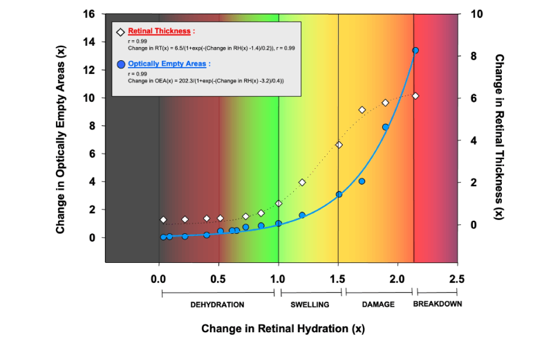
The study team took 6.0 mm sections of the human sensory retina excised from 18 fresh (<24 hours) donor eyes. The sections were either exposed to various osmotic stresses between 90 and 305 mOsm or dehydrated under a laminar flow hood. Change in tissue weight was used to calculate the retinal water content (RWC). Image analyses were conducted on OCT between 0 and 180 minutes to assess RT and “optically empty areas” (OEAs) representing intraretinal fluid. Correlations were sought among RWC, OEA, and RT. The effect of RH on retinal cell viability (RCV) was assessed with the Live-Dead Cell Viability Assay.
“Our findings showed a strong correlation between retinal fluid content and time, indicating that the exponential decay model (a fixed fraction of the element will decay in each unit of time) accurately demonstrates the relationship between these two variables,” says Dr. Tezel. “We observed a similar correlation between optically empty areas and retinal thickness measurements in relation to time.”

Change in OEAs in the left y-axis and change in RT in the right y-axis, compared to RH values in the x-axis.
Developing a new algorithm that predicts the behavior of intraretinal fluid over time
Using the data and correlations from the study, the team developed a unique algorithm that predicts the behavior of intraretinal fluid over time, providing valuable insights into the dynamics of retinal tissue under varying hydration conditions.
“This algorithm allows us to calculate the amount of water accumulated in the retina and its movement,” says Dr. Tezel. “Considering that all major blinding retinal diseases, such as wet age-related macular degeneration, diabetic eye disease, and retinal vein occlusions, cause blindness mainly by fluid leaking into the retina, estimating the amount of fluid using this algorithm will have implications in gauging the efficacy of the different treatment regiments and predicting the prognosis of the patients.”
“With this algorithm,” he continues, “we will be able to sharpen our decision-making process and know which patients will benefit from treatment and which ones will not. Knowing when to treat is also important, because if a patient seems fine at the time of the evaluation, we should not treat because there’s no reason to risk the person’s eye with unnecessary treatments.”
With this algorithm, we will be able to sharpen our decision-making process and know which patients will benefit from treatment and which ones will not. Knowing when to treat is also important, because if a patient seems fine at the time of the evaluation, we should not treat because there’s no reason to risk the person’s eye with unnecessary treatments.
— Dr. Tongalp Tezel
The study team views the algorithm as bridge to a future when artificial intelligence (AI) can be utilized to aid in clinical decision-making. “We are entering the exciting area of AI with these correlations and algorithms,” says Dr. Tezel. “With the power of AI, we will have the ability to become more precise in diagnosing disease entities, knowing when to interfere, and making an accurate prognosis.”
We are entering the exciting area of AI with these correlations and algorithms. With the power of AI, we will have the ability to become more precise in diagnosing disease entities, knowing when to interfere, and making an accurate prognosis.
— Dr. Tongalp Tezel
“This is the new world of precision medicine, a new horizon in care where we are getting more precise and more delicate in our analyses,” he adds. “With the aid of this algorithm and enhanced with AI, the information we capture from patients will be much more meaningful to help us determine the best care for each individual patient, and, in turn, allow us restore vision in a wider patient population.”


