Head and neck squamous cell cancer is the sixth leading cause of cancer and cancer-related deaths in the world, with an estimated 562,000 people diagnosed and some 278,000 who died from the disease in 2020 according to Cancer.Net®. In the United States, head and neck cancer accounts for about 4 percent of all cancers, with more than 66,000 newly diagnosed cases and approximately 15,000 deaths expected in 2022.
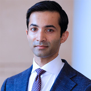
Anuraag S. Parikh, MD, is a head and neck surgeon in the Department of Otolaryngology – Head and Neck Surgery at NewYork-Presbyterian/
Dr. Anuraag Parikh
“Despite advances in surgery, radiation, and chemotherapy, as well as the development of immunotherapy over the past decade, outcomes in head and neck cancer, particularly at the advanced stage, have shown only modest improvements,” says Dr. Parikh. “This has been a main motivating factor for me to pursue research at a biological level. Very few biomarkers in this patient population are in clinical use except for human papilloma virus status, which is the only biomarker that is of clinical utility in head and neck cancer at this point. While a number of large-scale sequencing studies have begun to define pathways that are active and mutations that are present in these cancers, the important takeaway has been that head and neck cancer is driven by an accumulation of mutations in tumor suppressor genes. These are much more difficult to target than oncogenes, which are typically not mutated at a high rate in head and neck cancer.”
“These sequencing studies, however, tend to overlook a concept called tumor heterogeneity,” continues Dr. Parikh. “Traditionally, tumors have been thought to arise from a single cell that gained the ability to invade and proliferate unchecked and out of control. The tumors were considered to be clonal in which all the cells looked like one another and arose from this single cell. More recently, it has become clear that tumors are in fact made up of a variety of different cells, including non-malignant cell types such as fibroblasts, endothelial cells, immune cells, and so on, as well as a diversity of malignant cells. The greater the intratumoral heterogeneity that is present, the poorer patients tend to do.”
In the July 15, 2022, online issue of Cancer Cytopathology, Dr. Parikh and Sidharth V. Puram, MD, PhD, a head and neck surgeon at Washington University, describe how single-cell sequencing technologies, including methods to interrogate the single-cell transcriptome, have provided a more nuanced understanding of intratumoral heterogeneity in HNSCC as well as an appreciation of intratumoral cellular subpopulations. But an optimal approach to identify biomarkers that can predict survival outcomes and guide treatment for head and neck cancers has yet to be revealed. During his training at the Massachusetts Eye and Ear Infirmary, Dr. Parikh and his colleagues there utilized single cell RNA-seq to characterize head and neck squamous cell carcinoma and uncovered a potential role of partial epithelial-to-mesenchymal transition (p-EMT) warranting further inspection. They went on to demonstrate the association of p-EMT in primary tumors with the presence of nodal metastasis, suggesting that p-EMT biomarkers may aid in surgical decision-making and/or adjuvant therapy planning.
“From a biological standpoint, we are now looking to understand the role of a subpopulation of cancer cells undergoing p-EMT, which we identified previously in our single-cell sequencing efforts, with modeling in a three-dimensional organoid,” says Dr. Parikh. “By modeling the biology and the functional impact of this particular pathway and understanding its therapeutic susceptibilities, we seek to describe how targeting this pathway may impact invasion and metastasis.”
Transitioning from Single Cell Sequencing to 3D Organoid Modeling
“Traditional in vitro modeling of cancers has been done with cell lines from patient tumors grown in a Petri dish. However, there are multiple challenges with these two-dimensional cell-line models,” explains Dr. Parikh. “One is that it is hard to derive them from patient samples with a high success rate. Additionally, they do not adequately capture the heterogeneity of cancer cells that are present within tumors.”
Organoids are essentially cells grown in three-dimensional structures embedded within a matrix that mimics the basic membrane of the in vivo setting. “This allows us to capture a much greater diversity of cells and also capture intercellular interactions and differentiation gradients in a way that two-dimensional cell cultures do not,” notes Dr. Parikh. “Furthermore, 3D organoids can be derived from patient tumors at a success rate in excess of 80 percent, a much higher success rate than achieved with 2D cell lines. This means that we can almost reliably model the tumors of every single patient who comes in the door.”
Using 3D organoid models, we are aiming to develop better models of tumor heterogeneity and particularly in rare tumor subpopulations that may be functionally and prognostically relevant.
— Dr. Anuraag Parikh
Dr. Parikh is guided in his organoid work by his mentor and scientific colleague, Hiroshi Nakagawa, MD, Associate Professor of Medicine and a member of the Herbert Irving Comprehensive Cancer Center at Columbia. Dr. Nakagawa conducts research with unique model systems on the fundamental mechanisms in epithelial renewal, differentiation, and cell fate determination in esophageal diseases as well as cancers of the adjacent oropharyngeal mucosa. While on the faculty at the Perelman School of Medicine, University of Pennsylvania, Dr. Nakagawa served as a member of the multicenter research team that described for the first time the successful generation and characterization of tumor-derived 3D organoids from patients with esophageal and oropharyngeal squamous cell carcinomas. The study demonstrated “their utility as a robust platform to analyze cancer cell heterogeneity, evaluate therapy response, and explore therapy resistance mechanisms with the potential of translation in the setting of personalized medicine.” (Cellular and Molecular Gastroenterology and Hepatology, 2018 Sep 14)
Ultimately, Dr. Parikh hopes to be able to reliably generate organoid models from every single patient's tumor and create a bank of targeted agents for the different pathways that may be active in head and neck cancer. “This could make it possible for us to be able to test individual patient's tumors for responses to therapies that are not widely used in head and neck cancer,” says Dr. Parikh. “We still have a fair bit of characterization and patient stratification to do, but a critical step that we are trying to achieve now is developing a model system that mimics in vivo biology much better than traditional existing model systems such that we can reliably predict a patient’s tumor response.”
“As part of the larger equation, we hope this type of model will help us understand the viability of a number of different chemotherapies and targeted therapeutics across patients with head and neck cancer,” says Dr. Parikh. “Modeling the impact of, for example, radiation therapy is also something that we can do with organoids, which have been shown to recapitulate response to radiation consistent with response seen in patients. The most attractive feature of the 3D organoid model system is how easily and reliably it can be generated from a patient's tumor in about a week. This model could conceivably fit into the workflow of deciding between alternate treatments for a given patient.”
Expanding Organoid Modeling Beyond HNSCC
Dr. Parikh and his Columbia colleagues are also evaluating the potential of organoids to modeling tumor biology in adenoid cystic carcinoma, a rare and deadly salivary gland tumor. They are looking to develop a similar 3D organoid modeling of tumor heterogeneity in adenoid cystic carcinoma that may lead to therapeutic susceptibility. “The same traditional 2D cell-line models, which were not very good for mucosal squamous cell cancers, do not even exist for adenoid cystic carcinoma,” says Dr. Parikh. “Therefore, adenoid cystic carcinoma presents a unique challenge that may be overcome by generating 3D organoid models that would allow us to better understand potential targeted therapies in these tumors.”
With a focus on the long-term, big-picture goal, Dr. Parikh believes that utilizing organoids essentially as avatars of patient tumors for the sake of personalized medicine is very much within reach. “I expect that the next 5 to 10 years will be focused on leveraging 3D organoids to uncover an understanding of intratumoral heterogeneity and how they play a role in the biology of tumors across patients. Subsequent to this, the model system will be ripe for a personalized medicine approach. Here at Columbia, we are well-poised to pioneer the application of this technology to understand tumor biology, expose novel therapies, and discover ways to optimize treatment of patients with head and neck cancers.”


