


While the most concerning complication of COVID-19 is acute respiratory failure, cardiovascular damage from the virus is also commonly seen in some 20 to 30 percent of hospitalized patients. Acute cardiac injury has been shown to be associated with a significantly greater mortality rate in COVID-19 patients than other risk factors, including age, chronic pulmonary disease, or history of cardiovascular disease.
Seeking to understand the cause behind the cardiac damage, researchers co-led by faculty at Weill Cornell Medicine, Columbia University Vagelos College of Physicians and Surgeons, and others, conducted a study of heart samples of patients with COVID-19 that revealed abnormal inflammation of macrophages in the heart tissues. In the study, published on June 25, 2021, in Circulation Research, the team established an immuno-host coculture system to examine the macrophage-induced host cell damage following SARS-CoV-2 infection. They also used the system to identify two existing FDA-approved drugs that may be able to prevent or ameliorate this type of cardiac complication in patients with COVID-19.
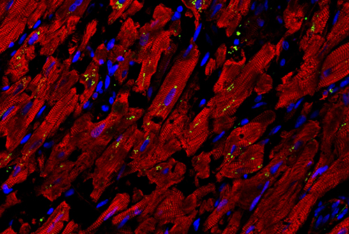
Confocal image of heart autopsy sample of a COVID-19 patient. The image showed the damaged structure of cardiomyocytes (red) and increased expression of chemokine, CCL2 (green), which recruits macrophages that further damage heart tissue.
Credit: Dr. Liuliu Yang
The research team included Shuibing Chen, PhD, Kilts Family Associate Professor of Surgery and Associate Professor of Chemical Biology in Surgery; Todd Evans, PhD, Associate Dean for Research; Peter I. Pressman, MD, Professor in Surgery; Robert E. Schwartz, MD, PhD, Associate Professor of Medicine in the Division of Gastroenterology and Hepatology, Weill Cornell Medicine; and virologist David D. Ho, MD, Professor of Microbiology and Immunology at Columbia University Vagelos College of Physicians and Surgeons. They noted that prior studies of COVID-19 pathology used animal models, but an advantage of their model is that it can use human-derived cells and be adapted to screen for potential therapies.
Dr. Shuibing Chen

Dr. David Ho
In the study, the team compared autopsy samples from non-COVID-19 donors and COVID-19 patients using RNA sequencing and immunohistochemistry and observed increased expression levels of CCL2 (C-C motif chemokine ligand 2) and macrophage infiltration in heart tissues of the COVID-19 patients.
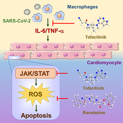
Dr. Chen and Dr. Evans pooled their expertise in cell-based disease models and engineered a lab-dish culture of human pluripotent stem cell-derived cardiomyocytes and macrophages. They found that adding the SARS-CoV-2 virus led to infection of the cardiac cells, secretion of CCL2 from the infected cells, and a resulting increase in macrophage inflammatory activity. The virus did not infect the macrophages themselves, but in their inflammatory state they released highly reactive and damaging molecules that triggered the death of many of the cardiac cells.
Based on their recently published data, the researchers believe that infection with the SARS-CoV-2 virus directly injures and kills cardiac cells. In the meanwhile, the infected cardiac cells secrete chemokines to recruit monocytes, which change into macrophages when they infiltrate the heart. The macrophage likely helps to remove infected and dying cells. Their new study suggests that when macrophages go into a hyper-inflammatory mode, the damage they cause can account for a large part of the pathology seen in autopsied hearts from COVID-19 patients.
(Source: Circulation Research)
They then used their COVID-19 model to search for potential treatments. Performing a high content screen of nearly 1,300 drugs already approved by the FDA, they found two compounds – ranolazine and tofacitinib – that robustly reduce the levels of reactive oxygen molecules and reduce the deaths of cardiac muscle cells. The team found evidence that ranolazine, which is used to treat angina, reduces the production of reactive oxygen molecules. They also learned that tofacitinib, used to treat rheumatoid arthritis and other serious inflammatory conditions, protects cardiac cells by inhibiting the Janus kinase pathway required in many aspects of immune activation. A recent clinical trial of patients hospitalized with COVID-19 pneumonia has shown that tofacitinib led to a lower risk of death or respiratory failure than placebo. A similar Janus kinase-inhibiting drug, baricitinib, was also shown in a clinical trial to shorten recovery times for hospitalized COVID-19 patients, and was approved by the FDA in November for emergency use in such cases.
The investigators hope that the potential treatments identified can be tested in large-scale clinical trials and they plan to continue to develop cell-culture models for the heart and other organ systems affected by COVID-19.