Pancreatic Cysts: When is There a Cause for Concern?

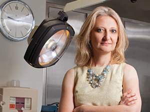
Dr. Tamas A. Gonda
With the use of advanced abdominal imaging techniques, the incidental identification of pancreatic cysts is becoming increasingly more common. “Pancreatic cysts are the most readily identifiable precancerous lesions for pancreatic cancer,” says Tamas A. Gonda, MD, Director of Endoscopic Research in the Division of Digestive and Liver Diseases at NewYork-Presbyterian/Columbia University Irving Medical Center, and a member of Columbia’s Pancreatic Cyst Surveillance Program. “As such, they represent an opportunity for us to identify patients who are at risk for developing pancreatic cancer so that hopefully we can intervene at a time when we can prevent a progression to invasive disease.”
While cysts represent precancerous lesions, they are also very common, with estimates between five and 20 percent of people in the United States having pancreatic cysts. “Obviously, that’s a huge number, and pancreatic cancers are nowhere near that high,” says
Although the majority of pancreatic cysts are benign, mucinous cysts represent at least a third of all cysts and are capable of malignant transformation. Mucinous cysts include mucinous cystic neoplasms and intraductal papillary mucinous neoplasms.
Understanding the Variables in Risk Stratification
“Much of our work is focused on understanding when a patient walks through the door if he or she is likely to be more of a high-risk person versus a low-risk person,” says Dr. Gonda. This requires an integrated and truly multidisciplinary approach with active participation of surgeons, radiologists, and gastroenterologists. Beth A. Schrope, MD, PhD, Surgical Director of the Pancreatic Cyst Surveillance Program in the Pancreas Center at Columbia, and Dr. Gonda work closely in pursuing aggressive investigations on how to best stratify an individual’s risk of developing pancreatic cancer. The first step is to examine demographics and patient characteristics. Patients undergo a comprehensive protocol that enables the physicians to capture as many variables as possible to better understand the risks of cancer and counsel them regarding the possible treatment options, including surgery.

Dr. Beth A. Schrope
“Columbia is now participating in several international multicenter studies that are focused on collecting data on a large number of patients with a very large number of variables,” says Dr. Gonda. “This is to help us identify what the real risk is for a patient to develop any kind of worrisome cyst. We ask if there is a personal history of other types of cancers, or the presence of associated pancreatitis, and gather detailed information about their family history of cancer. We are also beginning to recognize that metabolic parameters, such as diabetes, may be a potential marker of somewhat elevated risk.”
The next phase in the risk stratification algorithm involves radiologic imaging, an area that Dr. Gonda and his colleagues have researched at length. “Patient characteristics combined with our expertise in the radiographic characterization of pancreatic lesions provide us with a decent sense of the type of cyst we are facing.”
Patients in the Pancreatic Cyst Surveillance Program are given an informed evaluation of their risk and recommended next steps based upon the physicians’ findings. “Twenty to 40 percent of patients will require further investigation and more invasive studies,” says Dr. Gonda. “First we perform an endoscopic ultrasound with detailed imaging to evaluate the cystic lesion. Several new methodologies allow us to image the cysts in greater detail than ever before possible. Endoscopic ultrasound with confocal laser endomicroscopy enables us to obtain images from the cyst wall. The newest innovation is an endoscopic ultrasound-guided direct biopsy of the cyst wall letting us, for the first time, to obtain tissue from these cystic lesions.”
“The most precise, personalized imaging technologies will rely on highly specific molecular markers that can be used for imaging targets,” says Dr. Gonda. “We are still at the stage of using non- individualized imaging modalities to stratify risk. Then we obtain biopsy-based information to identify the one or two highly specific markers that can then be specifically imaged. But taking a biopsy of a pancreatic cyst is more challenging than your typical biopsy because what you usually access in pancreatic cysts is fluid, not cells. That is why we need to turn to complex molecular analysis of the tissue to help us distinguish the risk potential of a lesion. If you can identify a molecular marker that can be detected, then you have an amazing imaging tool to scan the cyst and determine whether or not a particular molecular abnormality exists. We are coming full circle on personalizing this approach, but we are not there yet.”
Dr. Gonda stresses that a high-volume, specialized center such as the Pancreatic Cyst Surveillance Program at Columbia allows for the expertise and input from every member of the team. “Our radiologists are highly in tune to identifying and utilizing the newest imaging technologies. Our pathologists are highly in tune to the nuances of the questions we are asking of tissue samples. And our expertise as endoscopists in cutting-edge imaging and biopsy technology allows us to minimize the number of procedures and interventions that patients undergo. Our hope is to be able to identify the relatively small subset of patients who can be saved by a preventive intervention.”
To this day, the most impactful intervention to prevent pancreatic cancer from developing in pancreatic cysts is surgical resection. Surgical innovation over the last one to two decades has revolutionized pancreatic surgery. According to Dr. Beth Schrope and John A. Chabot, MD, Executive Director of the Pancreas Center, “Our surgeries are much safer and our outcomes are better than before. We are using increasingly less invasive approaches, including laparoscopic and robotic surgery, for the majority of patients with cystic neoplasms. These procedures allow greater oncologic precision and faster recovery. Nonetheless, the goal is to develop diagnostic and nonsurgical techniques to minimize the need for resection in the majority of patients.”
Reference Articles
Gausman V, Kandel P, Van Riet, PA, Kayal M, Do C, Poneros JM, Sethi A, Gress FG, Schrope BA Luk L, Hecht E, Jovani M, Bruno MJ, Cahen D, Wallace MJ, Gonda TA. Predictors of progression among low-risk intraductal papillary mucinous neoplasms in a multicenter surveillance cohort. Pancreas. 2018. [In press]
Xu MM, Yin S, Siddiqui AA, Salem RR, Schrope B, Sethi A, Poneros JM, Gress FG, Genkinger JM, Do C, Brooks CA, Chabot JA, Kluger MD, Kowalski T, Loren DE, Aslanian H, Farrell JJ, Gonda TA. Comparison of the diagnostic accuracy of three current guidelines for the evaluation of asymptomatic pancreatic cystic neoplasms. Medicine (Baltimore). 2017 Sep;96(35):e7900.
Kayal M, Luk L, Hecht EM, Do C, Schrope BA, Chabot JA, Gonda TA. Long-term surveillance and timeline of progression of presumed low-risk intraductal papillary mucinous neoplasms. American Journal of Roentgenology. 2017 Jun 7:1-7.
Related Publications


