


With the number of COVID-19 cases in the United States on the decline albeit with a close watch for emerging variants, data on long-term effects on patients who had the virus continue to surface. While some of the reports on enduring health concerns are anecdotal; a comprehensive body of research is building as clinicians and scientists around the world apply their expertise to studying post-acute sequelae of SARS-CoV-2 infection.

Dr. Claire McGroder, Lead Author
With chronic respiratory problems chief among the persistent conditions, pulmonology and critical care medicine specialists are key investigators in the effort to better understand the ongoing impact of COVID-19 on lung function and disease. Among them are Christine Kim Garcia, MD, PhD, Chief of the Division of Pulmonary, Allergy, and Critical Care Medicine at NewYork-Presbyterian/
Following the 2003 outbreak of SARS-CoV-1, studies pointed out there were persistent chest imaging abnormalities and histopathological findings of lung fibrosis in a majority of the survivors. With this in mind, the Columbia research team, which also included radiologists, Mary M. Salvatore, MD, and Belinda M. D’Souza, MD, undertook a study to determine if the current SARS virus is also associated with worse fibroproliferative response than other pneumonias. Dr. McGroder, a pulmonary specialist in interstitial lung disease who recently joined the Columbia faculty, served as the study's lead author.
Specifically, their goal was to characterize associations of pulmonary radiographic and physiologic sequela of severe COVID-19 and identify independent risk factors for the development of post-COVID fibrosis. The prospective cohort study involved 76 patients hospitalized with severe COVID-19 at NewYork-Presbyterian/
Four months following hospitalization, the patients underwent a non-contrast high resolution chest CT and pulmonary function testing, as well as measurement of 6-minute walk distance, assessment of the frailty phenotype, and a blood draw to determine telomere length of genomic DNA. Radiographic patterns were classified into a non-fibrotic or fibrotic group.
The findings of the study, published in the December 2021 issue of Thorax, demonstrated:
- Fibrotic-like abnormalities were more common in patients who were mechanically ventilated (72 percent) compared with those who were not (20 percent)
- Ground glass opacities (43 percent) were the most common radiographic abnormality followed by reticulations (39 percent) and traction bronchiectasis (28 percent)
- Ground glass, reticulations, and traction bronchiectasis scores correlated with a reduction in pulmonary function
- Greater severity of illness at hospitalization, a longer duration of mechanical ventilation, and shorter blood leucocyte telomere length were independent risk factors for the development of fibrotic-like abnormalities
Short telomere length has been shown to be a risk factor for the development of idiopathic pulmonary fibrosis (IPF). In this study, longer telomere length appeared to be protective and shorter telomere length appeared to be a risk factor for the development of fibrosis-like abnormalities following severe COVID-19.
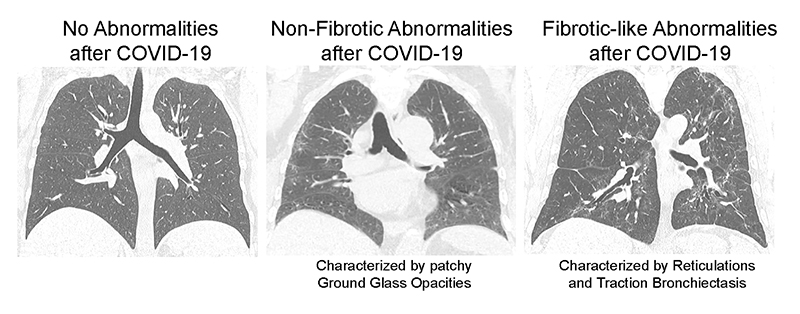
Representative CT chest scans demonstrate no abnormalities (left), non-fibrotic abnormalities (center) and fibrotic-like abnormalities (right) after severe COVID-19. All three panels show a representative coronal section image from one of the study participants.
Additional results showed that participants had a number of deficits in function as follows:
- 53 percent had a reduced diffusion capacity
- 78 percent had a decreased 6-minute walk distance
- 18 percent remained more than 10 percent below their baseline weight
- 53 percent had weak grip strength
In summary, the authors note that the “study reveals significant respiratory symptoms and morbidity associated with severe COVID-19.” Survivors of severe COVID-19, with and without mechanical ventilation, showed fibrotic-like radiographic abnormalities four months after being hospitalized and that these radiographic abnormalities correlated with reduced lung function, cough, and frailty. Importantly, this is the first study to identify age-adjusted telomere length as an independent risk factor for the development of post-COVID lung fibrosis.
