


New Horizons in Hematologic Oncology
- AML: An Unprecedented Potential
for Pre-Cancer Interception - Activating & Abrogating
Lymphoma Development - Adoptive Immunotherapy
with Cord Blood Cells - Bone Marrow Transplantation
Redeems its Pledge - A New Era in Treatment
of Sickle Cell Anemia - CAR T-Cells
Therapy & Beyond - An In-House Immune Cell
Production Laboratory - Circulating Tumor DNA:
Measurement for Optimizing Therapy - Supporting Up &
Coming Scientists
AML: An Unprecedented Potential for Pre-Cancer Interception
Acute myeloid leukemia (AML) is an aggressive myeloid malignancy characterized by clonal expansion of hematopoietic precursors in the bone marrow that results, ultimately, in bone marrow failure. Although there have been recent advances in the treatment of AML, the disease continues to carry a poor prognosis. While certain familial, genetic, and environmental factors may increase an individual’s predisposition to the disease, the cause remains unknown for the vast majority of patients — making early detection or disease interception strategies virtually impossible to consider ... until now.

Research by Weill Cornell Medicine faculty on identifying the potential for preventing the development of AML was the cover story in the July 9, 2018, issue of Nature Medicine. The cover art was created by Weill Cornell faculty.
The cover is reprinted here with permission from Springer Nature.
A study by Weill Cornell investigators, led by Pinkal Desai, MD, is the first to describe a genomic pre-malignant landscape of acute myeloid leukemia. The research, published in the July 9, 2018, issue of Nature Medicine, and the issue’s cover feature, found that acquired mutations in high risk genes could be identified in the peripheral blood of a group of patients up to a decade before AML was finally diagnosed.
“ Our findings provide an unprecedented opportunity to start thinking about ways to intercept AML to delay or prevent it before it develops in patients at risk.”
&nmdash; Gail J. Roboz, MD
The researchers evaluated blood samples from participants in the Women’s Health Initiative (WHI), a major multicenter observational and clinical study by the National Institutes of Health that began in 1991. The WHI followed more than 100,000 women for many years, enabling the establishment of a unique and clinically annotated database. Through this database, the Weill Cornell Medicine researchers were able to identify subjects who developed AML and match them to subjects within the population studied who did not develop AML. Using very deep next-generation sequencing technology, they identified certain mutations and patterns of mutations that arose in subjects who developed AML nearly a decade before they were eventually diagnosed with the disease.
Specifically, the Weill Cornell team was looking for trace levels of mutations in genes associated with leukemia in blood from about 200 women who later developed the disease. They then compared the results with approximately 200 women in a control group who remained healthy. The study found that 69 percent of women in the AML group had mutations in one or more of the genes examined, whereas only 31 percent in the control group had mutations associated with the disease.
Participants with mutations in IDH1, IDH2, TP53, DNMT3A, TET2, and spliceosome genes were significantly more likely to develop AML compared to the control group. The researchers also discovered that the number, pattern, and burden of different mutations were associated with varying levels of risk for developing AML and with the timing of when the disease emerged. Notably, all of the women in the cohort who had mutations in IDH1, IDH2, or TP53 eventually developed AML. The research team is now working on strategies on how to identify at risk populations and follow them prospectively.

Gail J. Roboz, MD , Director, Clinical and Translational Leukemia Program, Weill Cornell Medicine
The researchers emphasize, however, how challenging it is to figure out who should be screened for these mutations and, importantly, that not every individual with IDH or other AML-related mutations will go on to develop the disease. One reason is clonal hematopoiesis of indeterminate potential, or CHIP, a common aging-related phenomenon. It is well recognized that certain somatic mutations are present in patients as a normal part of aging and will not develop into a malignancy. The question then becomes, “How is the discovery applied to patients with a higher risk than normal of developing AML?”
The researchers suggest that patients with familial predisposition syndromes, and those who have had chemotherapy or radiation exposure, are going to be important groups to target for prospective monitoring. But there are still questions as to why some people with these exposures develop AML and others don’t. The researchers are continuing their work to identify other high risk groups for the development of hematological malignancies within the Women’s Health Initiative and are also initiating studies to determine if monitoring the pattern and burden of somatic mutations over time will lead to early intervention strategies.
While the researchers and scientific community consider this groundbreaking work and anticipate that it will lead to clinical trials of potential disease interception strategies in the future, they are quick to point out that healthy individuals should absolutely not rush to be randomly screened for AML-related mutations as the research is not at the point of being able to provide guidance on interventions.
Activating and Abrogating Lymphoma Development
New studies from Weill Cornell Medicine researchers have revealed the importance of two gene-regulation proteins — LSD1 and TET2 — in the development of lymphomas. Most lymphomas develop from B cells and are likely to arise during a distinct phase of the B cell life cycle in which the cells form germinal centers. B cells in germinal centers proliferate rapidly and deliberately destabilize their genomes to cause mutations to their antibody genes. The changes in B cells that put them into this state of fast growth and fast mutation also raise the risk that they will turn cancerous, the reason germinal center B cells are a major focus of lymphoma research.
LSD1: In the Beginning
The protein LSD1 plays a major role in starting and sustaining lymphomas. In a paper published in the December 10, 2018, issue of Nature Immunology, Weill Cornell researchers examined the function of LSD1 in germinal center B cells. The protein works to remove small chemical marks called methyl groups from histones, which associate with the genome. Methyl groups and other related chemical marks serve as switches, or “epigenetic marks,” that regulate genes. When LSD1 proteins become numerous and active in a cell, they can remove many methyl groups across the genome, thus broadly reprogramming the activity of the cell.
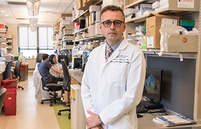
Ari M. Melnick, MD, Gebroe Family Professor of Hematology/ Oncology, Weill Cornell Medicine
The scientists found that LSD1 is produced in B cells when they are activated by the immune system and is required to encode the genetic programming that enables B cells to form germinal centers. They also revealed that LSD1 induces its effects through close partnership with BCL6, a protein that had already been linked to germinal centers as well as to lymphomas. In fact, in mice with lymphoma driven by BCL6 activity, removing LSD1 greatly slowed the tumor growth. The researchers are now working on a therapeutic strategy to counteract this effect of LSD1 in B cell lymphomas.
TET2: The End Game
In collaboration with several TET2 laboratories, Weill Cornell researchers examined not the beginning but the end of the germinal center phase of B cell development. The scientists found that TET2, another protein that alters epigenetic marks on DNA, is required to shut off the germinal center reaction in B cells and direct the cells into the next stage of their life cycle where they are far less likely to turn cancerous.
The study clarified that TET2 is mutated in many cases of B cell lymphoma because germinal center B cells lack a working TET2 protein that blocks B cells from exiting the cancer-prone germinal center state. The damaging effects of TET2 loss, however, can be addressed using a recently developed class of drugs called HDAC3 inhibitors, which may restore the protective effect of TET2 in tumors where it has been mutated. In their experiments so far, the researchers have found that lymphomas that arise from the lack of TET2 appear to be highly sensitive to HDAC3 inhibitors. These results advocate for sequencing TET2 in patients with lymphoma and for the testing of epigenetic therapies to treat these tumors. They hope to begin clinical trials of these drugs in selected patients with lymphoma in 2019. The results of the study were published in the October 1, 2018, issue of Cancer Discovery.
Adoptive Immunotherapy with Cord Blood Cells
Patients with relapsed or refractory acute myelogenous leukemia have limited treatment options and a generally poor prognosis. An early study by researchers at Weill Cornell and at the National Cord Blood Program has shown encouraging results on the use of umbilical cord blood cells and salvage chemotherapy to induce remission in patients with very advanced AML or myelodysplastic syndrome (MDS), which can serve as a bridge to allogeneic hematopoietic stem cell transplantation (HSCT). Others have used cells from partially related family donors for the same purpose. But according to the Weill Cornell researchers, umbilical cord blood cells have two advantages. They are readily available through blood banks to treat thereby avoiding the complicated logistics required for haploidentical donors, and there is ample evidence that umbilical cord blood cells have more powerful antileukemic effects than cells from adult donors.
In their study, the researchers prospectively evaluated the feasibility and tolerability of infusing cord blood units (CBUs) selected to have specific alloreactive properties, and administered after salvage chemotherapy, to 21 patients with refractory AML or MDS treated at NewYork-Presbyterian/Weill Cornell. All CBUs were obtained from the National Cord Blood Program. The unrelated CBUs were infused within 72 hours after completion of the chemotherapy regimen and no sooner than 24 hours after administration of the last dose of chemotherapy.
Response to treatment was defined as effective cytoreduction, i.e. <10 percent residual blasts in a hypocellular bone marrow or no blasts in an acellular bone marrow obtained approximately 14 days after CB cell infusion. Overall, 12 of the 21 patients proceeded to HSCT at a median of 44 days after cell therapy. Ten patients had effective cytoreduction, including four with residual bone marrow blasts <10 percent and six without residual blasts. Although treatment-related adverse events were frequent, they were in line with what would have been expected from salvage chemotherapy alone, and few were attributed to the CBU infusion.
Koen van Besien, MD, PhD, Director, Stem Cell Transplant Program, Weill Cornell Medicine
The findings, reported first in the November 9, 2018, online edition of Biology of Blood and Marrow Transplantation: Journal of the American Society for Blood and Marrow Transplantation, demonstrated that the combination approach salvage can induce disease control, can serve as a bridge to allogeneic HSCT, and has an acceptable incidence of adverse events. Furthermore, the potential for alloreactivity can be enhanced through the selection of CBUs targeting shared IPA and/or NIMA matches with the recipient.
The authors concluded that additional prospective studies are needed to establish efficacy in AML, MDS, and possibly in other hematologic malignancies, and also to identify recipient and CBU-related predictors of response and of toxicity.
Bone Marrow Transplantation Redeems its Pledge
Bone marrow transplantation has long held great promise as a definitive cure for certain hematopoietic malignancies or benign hematologic conditions (e.g. sickle cell disease). In fact, in 1957, Columbia physicians were among the first to perform a bone marrow transplant in humans. Furthermore, dating back more than 70 years, hematopoietic cells had been shown to have the potential to induce true immunologic tolerance (i.e. lack of rejection or graft acceptance without immunosuppression), which is considered the Holy Grail in transplantation medicine as it would obviate the need for lifelong treatment with medications to suppress the immune system. Additionally, it has been known that the response of the donor immune system against the recipient can be potentially harnessed against the underlying malignancy (graft-versus-leukemia [GVL] effect). Despite significant progress over the last decades, allogeneic transplantation is still associated with potentially life-threatening complications like graft-versus-host disease (GVHD), infectious complications, and recurrence of the underlying malignancy due to failure of the GVL response. These risks have precluded widespread application of transplant and limited its use for the treatment of benign conditions.
Markus Y. Mapara, MD, PhD, Director, Blood and Marrow Transplantation and Cell Therapy, NewYork-Presbyterian/Columbia
Recent developments in targeted therapy or drug discovery and revolutionary advances in genetic engineering of stem and immune cells have now ushered in a new era in cell therapy that may allow the expansion of BMT and cell therapy options to a much broader group of patients. The BMT and Cell Therapy program at Columbia is at the forefront of these research developments and is offering these treatment modalities to their patients. The research efforts at Columbia focus on the development of novel approaches to treat and prevent GVHD; the cure of sickle cell disease using donor transplants and genetic engineering of patients’ own stem cells; cellular immunotherapy of cancers using genetically engineered T-cells, such as CAR T-cells; and in-house development of T-cells directed against viral diseases and leukemias.
“Targeting the JAK1/STAT1 pathway is one of the most exciting new approaches to treat or prevent graft-versus-host disease without blunting the anti-malignancy or graft-versus-leukemia effect in patients with blood cancers.”
— Markus Y. Mapara, MD, PhD
Graft-versus-host disease is one of the most common and serious side effects of allogeneic bone marrow transplant. One area of active research by clinician-scientists at Columbia is to mitigate the risk for GVHD while preserving the graft-versus-leukemia (GVL) effect and maintaining the curative potential of allogeneic transplant in cancer. These efforts are critical for the improvement of survival and quality of life of patients undergoing allogeneic transplantation and the Columbia research team has a track record in developing and implementing cutting-edge methods and bringing them from bench to bedside. Success of these efforts will allow allogeneic transplant recipients to live longer and without lifelong immunosuppressants.
By finding potentially druggable targets, the Columbia researchers could use a drug to suppress or prevent GVHD without blunting the anti-malignancy or GVL effect in patients with blood cancers. One signaling pathway they are particularly interested in is interferon/JAK/STAT1. Dr. Mapara’s group was the first to demonstrate that blocking the STAT1 pathway in donor cells could significantly reduce or even prevent GVHD. In his lab, researchers are currently trying to understand in more detail using mouse models how to better manipulate that approach and how this will affect the anti-tumor response in the human setting.
Taking the next step closer to clinical studies, the researchers are moving from using standard mouse models to working with humanized mouse models. The research involves a certain strain of mice that are highly immunosuppressed, which allows the investigators to establish human blood and immune systems in these mice. These experiments allow researchers to generate clear evidence that interferon signaling and the JAK/STAT pathway play a significant role, not only in mice, but also in the human immune system in terms of GVHD.
The initial mouse studies have been recently confirmed by early clinical trials, which demonstrated that drugs inhibiting the JAK1/2 molecule (which is upstream of STAT1) are active in patients with GVHD. Based on the Columbia studies and with the goal of further enhancing outcomes, the research team is now initiating a clinical trial that will focus on targeting JAK1 together with another key pathway, the IL-6 signaling pathway, in patients with steroid-refractory GVHD.
Eradicating GVHD is crucial because it would allow clinicians to also use bone marrow transplantation as an innovative strategy to assist solid organ transplant by inducing long-lasting and permanent immune tolerance. In close collaboration with the Columbia Center for Translational Immunology and the Kidney Transplant Program, a clinical protocol is open at Columbia in which patients receive a combined kidney and bone marrow transplant from the same donor. Building on the early success of this approach, the researchers are planning to add infusion of regulatory T-cells — which keep the immune system in check — following the combined kidney and bone marrow transplant to enhance the development of donor cell engraftment and tolerance induction. This second generation clinical trial will soon be ready to begin in patients.
A New Era in Treatment of Sickle Cell Anemia
Sickle cell disease caused by a single-gene defect in the beta-globin chain of hemoglobin has long been speculated to be an ideal target for gene therapy approaches. Furthermore, genetic engineering of autologous hematopoietic stem cells, if successful, would result in cure without the potential detrimental side effects of GVHD associated with allogeneic BMT. A true gene correction approach (i.e. swapping out the mutated beta-globin chain with a correct one) is not yet clinically available. However, other genetic approaches which focus on production of anti-sickling hemoglobin by a patient’s stem cells and which prevent the sickle hemoglobin to polymerize and induce vaso-occlusive crises show great promise. The Columbia BMT and Cell Therapy program is participating in two of these early stage trials which offer two different types of genetic engineering for patients with sickle cell disease.
The combined adult and pediatric BMT program at Columbia is one of the largest transplant programs for patients with sickle cell disease in the U.S. and offers the entire spectrum of allogeneic transplants using matched sibling, matched/mismatched unrelated to haploidentical donors. In line with the overarching interest of our program in eliminating GVHD together with our pediatric colleagues, we are conducting a trial in adult sickle cell patients using in vivo and ex vivo T-cell depletion to prevent GVHD in patients without matched siblings. This approach has proven to be very successful in preventing GVHD and thereby enables patients without a matched sibling to undergo this lifesaving procedure.
CAR T-Cells Therapy and Beyond
Since the first successful applications of CD19-targeting Chimeric Antigen Receptor (CAR) T-cells nearly a decade ago for adults with chronic lymphocytic leukemia and children with acute lymphoblastic leukemia, research in the field of cell therapy has soared, with several hundred clinical trials of CAR T-cells against multiple targets and other types of cell therapy approaches underway around the country. At Columbia, BMT faculty are studying the latest CAR T-cell therapies in clinical trials for aggressive lymphomas and other types of blood cancers, including indolent lymphomas, leukemias, and myeloma, as well as developing trials for difficult-to-treat solid tumors, such as pancreatic cancer, sarcoma, and melanoma.
While CAR T-cell therapy has had major success turning some incurable diseases into curable ones, there remains work to be done. CAR T-cells overcome one specific barrier in cancer treatment, which is target recognition, leading to dramatic responses initially but progression of cancer is still observed in some patients several months following therapy. Durable remissions, which are potential cures, range from 50 to 60 percent in patients with acute lymphoblastic leukemia and approximately 40 percent for patients with lymphoma. To address this, researchers at Columbia are in the early stages of developing approaches to increase the efficacy of existing CAR T-cells using combination therapies, for example, CAR T-cells with a checkpoint inhibitor, to help overcome barriers to treatment success, increasing response rates and durability.
Ran Reshef, MD, MSc, Director, Translational Research, BMT and Cell Therapy, NewYork-Presbyterian/Columbia
Columbia researchers also are exploring additional targets beyond CD19 and surface antigens. CAR T-cells are only able to identify and find extracellular proteins. However, a considerable number of tumor antigens do not present on the cell surface and are therefore unable to be identified. The Columbia team is now exploring alternative methods that would offer a greater ability to identify and attack cells that express intracellular antigens.
Cytokine release syndrome and neurological toxicity are the two most common toxicities associated with CAR T-cells and other forms of genetically modified T-cells. The BMT program has assembled a multidisciplinary team that includes experts in intensive care and neurology and have initiated research to understand the causes of cytokine release syndrome and neurotoxicity, and how to prevent or decrease these side effects. Currently, any patient undergoing CAR T-cell therapy at Columbia is seen by a neuroimmunologist prior to admission and undergoes a neurological assessment and brain MRI to obtain baseline information against which any changes can be compared following therapy. If a patient develops a severe neurological toxicity, a neurological ICU consult is immediately available. To date, the team’s experience in managing CAR T-cell side effects has been very positive, allowing the team to treat safely all types of patients, including patients with diseases that are highly refractory to standard treatments, as well as elderly individuals and those with comorbidities.
Pawel Muranski, MD, Director, Cellular Immunotherapy, NewYork-Presbyterian/Columbia
An In-House Immune Cell Production Laboratory
The immune system plays the central role in protecting against infections, but in recent years it also became clear that it is possible to generate cancer-fighting immune cells in the laboratory and to use them to completely eradicate tumors in patients. In 2017, Columbia University established a Cellular Immunotherapy Laboratory capable of producing immune cells under current Good Manufacturing Practice (cGMP) guidelines and meeting the FDA safety and quality regulations, making it one of the few institutions to have the ability to grow and manipulate T cells for therapy of patients with cancer or severe infections. Columbia researchers will now manufacture clinical grade immune cells based on the research taking place at Columbia, as well as in the labs of collaborating institutions. In the near future, the researchers are planning to implement the point-of-care in-house manufacture of CAR T-cells for treatment of lymphomas and leukemias in the lab Pawel Muranski, MD, Director, Cellular Immunotherapy, NewYork-Presbyterian/Columbia on campus. The researchers expect that this approach will make the lifesaving CAR T therapy significantly less costly and rapidly available to anyone in need. In addition, the scientists are using innovative methodologies, including gene engineering and other sophisticated manipulations, to generate novel potent immune cells called T helper cells targeting common cancers with the ultimate goal of evaluating their activity in clinical trials involving patients with advanced malignant diseases.
The Cellular Immunotherapy Laboratory is also developing T-cells for use as immune therapy and prevention of severe viral infections that frequently affect vulnerable patients with cancer and other diseases.
To that end, the researchers seek to obtain normal T-cells from healthy donors and to educate them to recognize the common viruses. In case of bone marrow transplant recipients, the protocol involves rebuilding the patient’s immune system immediately after the bone marrow transplant — the time of most vulnerability for the type of cells that will prevent the occurrence of infection. The researchers will also use a similar strategy as rescue treatment for many critically ill immunocompromised patients regardless of the underlying cause of infection, such as patients who received chemotherapy for cancer or immune suppressive drugs for autoimmune diseases or following solid organ transplantation.
Circulating Tumor DNA: A Measurement for Optimizing Therapy
One of the great challenges in oncology research is to develop therapeutic strategies that can overcome the ability of a tumor to evolve in response to treatment and thereby circumvent a successful outcome. Cancer cell populations undergo an evolutionary process that increases their diversity and enables them to acquire new mutations, resulting in a great deal of genetic diversity within each cancer. This process forces cancer therapies to target not a single disease but rather thousands of variations of disease within each individual patient, posing a significant therapeutic challenge.
In the Department of Hematology and Medical Oncology at Weill Cornell, researchers have been investigating cell-free circulating tumor DNA (ctDNA) — small fragments of DNA from dying tumor cells that are released into the bloodstream — for their potential role in detecting and following the progression of a patient’s tumor. Earlier work in chronic lymphocytic leukemia (CLL), published in Nature in 2015 by Dan Landau, MD, PhD, and colleagues while at Harvard, showed that if the disease relapses after therapy, the cancer genetically transformed to become more aggressive and treatment resistant. The emergence of promising targeted therapies, such as ibrutinib, for CLL provided clinicians with a new medication to treat patients with high-risk cancers or patients who experience a relapse, but scientists have already begun to document genetic changes in patients whose cancer progresses despite treatment with ibrutinib.
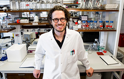
Dan Landau, MD, PhD, oncologist, Weill Cornell Medicine
By sequencing cancer samples from these patients more frequently and using computer modeling to predict the cancer’s genetic evolution, a team of scientists and collaborators from Weill Cornell Medicine, the National Heart, Lung and Blood Institute, Broad Institute, The University of Texas MD Anderson Cancer Center, among other institutions, were able to identify a subset of patients in whom cancer cells with certain genetic variations were dying less quickly than others. These changes could be seen as early as one to three months after treatment initiation, and well before the affected patients developed any signs of cancer recurrence. The investigators demonstrated that monitoring for genetic changes in CLL every month or two could identify early and ongoing changes in cancer genetics — even in patients who were responding to treatment — that were linked to the emergence of resistance and poor clinical outcomes.
A Role for Liquid Biopsy
The Weill Cornell researchers continue to investigate how tumor cells evolve to overcome therapy and expand this work to solid malignancies like lung cancer. One approach involves liquid biopsy, a highly sensitive platform for early detection of cancer through the measurement of tumor DNA fragments in a patient’s blood. Using sequencing information from DNA fragments obtained from patient plasma samples, a team of biologists, physician-scientists, data analysts, and engineers is looking broadly across the genome for relevant mutations to enable highly sensitive detection of cancer, even when it is not detectable by imaging tests such as CT scans.
The hope is that liquid biopsy will overcome current barriers to accurately diagnosing cancer when tumors are very small and only minute amounts of tumor DNA are available in the patient’s blood, but also optimize treatment by making it possible to continuously monitor genetic changes in cancer cells in real-time.
Supporting Up and Coming Scientists
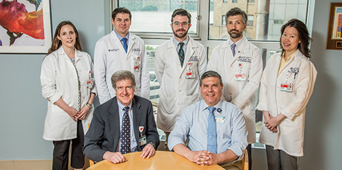
(Seated) Dr. Mark L. Heaney, Director of the Hematology/Oncology Fellowship Program, and Anthony Rainone, Fellowship Program Coordinator, NewYork-Presbyterian/Columbia (Standing) 2018 Young Investigator Award recipients: Dr. Ruth A. White, Dr. David H. Aggen, Dr. Daniel S. O’Neil, Dr. Shawn M. Sarkaria, and Dr. Jessica Yang
Five fellows in the Hematology/Oncology Fellowship Program at NewYork-Presbyterian/Columbia received a 2018 Young Investigator Award from the Conquer Cancer Foundation — a record number of awards for one institution. The highly prestigious and competitive award encourages and funds promising investigators during their transition from a fellowship program to a faculty appointment.
David H. Aggen, MD, PhD | Charles G. Drake, MD, PhD, mentor
Targeting Myeloid-Derived Suppressor Cells in Renal Cell Cancer with Combination lL-1 Beta and PD-1 Blockade
Dr. Aggen is developing novel immunosuppressive combination therapies for patients with kidney, bladder, and prostate cancer that target Interleukin 1. This cytokine, in combination with PD-1, is believed to drive the cancer cell and cause tumor immunosuppression.
Daniel S. O’Neil, MD, MPH | Alfred I. Neugut, MD, PhD, Paul Ruff, MBBCh, mentors
Quality of Breast Cancer Care in Five Public Hospitals in South Africa and Effects on Survival
As an epidemiologist interested in a global oncology research, Dr. O’Neill seeks to develop quality metrics appropriate for cancer centers in Africa and other low- and middle-income countries. His project calls on the extensive clinical and socioeconomic data available on breast cancer patients in the database of the University of Witwatersrand in collaboration with Columbia’s Mailman School of Public Health.
Shawn M. Sarkaria, MD | Gary K. Schwartz, MD, Lei Ding, PhD, mentors
Targeting PDGFRα-Expressing Stromal Cells in Primary Myelofibrosis
Dr. Sarkaria is studying primary myelofibrosis, a clonal stem cell-derived hematologic malignancy characterized by chronic myeloproliferation, atypical megakaryocytic hyperplasia, and bone marrow fibrosis. His research seeks to slow progression of myelofibrosis by targeting components of the tumor microenvironment to preserve normal bone marrow function and enhance drug efficacy.
Ruth A. White, MD, PhD | Timothy C. Wang, MD, mentor
Cholinergic Regulation of the Neural Microenvironment in Pancreatic Cancer
Dr. White is exploring the cholinergic regulation of the neural microenvironment in pancreatic cancer. She hopes that understanding biomarkers of the tumor microenviroment will lead to a targeted drug that will allow chemotherapies to get to the cancer cells more easily.
Jessica Yang, MD | Richard D. Carvajal, MD, mentor
Efficacy of Bromodomain and Extra-Terminal (BET) Protein Inhibition in Advanced Uveal Melanoma
Dr. Yang’s research focuses on novel therapies for rare and understudied tumor types and treatment strategies directed against specific oncogenic pathways common to different malignancies. In particular, the Young Investigator Award funds her work in developing clinical trials in advanced uveal melanoma.