


Improving Assessment of Anomalous Aortic Origin of the Coronary Artery
Evaluating anomalous aortic origin of a coronary artery (AAOCA), a rare congenital anomaly associated with sudden cardiac death in youth, is the focus of a recent study by cardiac surgeons and cardiology imaging specialists at NewYork-Presbyterian Morgan Stanley Children’s Hospital.

Dr. Emile Bacha

Dr. Kanwal Farooqi
“With more young athletes undergoing imaging studies to determine heart murmurs or other cardiac risks, we are seeing more diagnoses of anomalous aortic origin of a coronary artery,” says Emile Bacha, MD, Director, Congenital and Pediatric Cardiac Surgery, NewYork-Presbyterian Congenital Heart Center, and Chief, Cardiothoracic and Vascular Surgery at NewYork-Presbyterian/
With no specific guidelines for the surgical management of anomalous aortic origin of the right coronary artery, the pediatric heart team has been leading efforts to accurately diagnose this life-threatening condition. Patients are generally evaluated using a 2-dimensional echocardiogram or cardiac computed tomography. For the study, conducted under the leadership of Kanwal M. Farooqi, MD, a pediatric cardiologist with advanced training in imaging techniques, the researchers hypothesized that the high-risk anatomic characteristics would be more accurately assessed when presented in a 3D digital model.
In their study, published in the February 2021 issue of Annals of Thoracic Surgery, Dr. Farooqi and colleagues compared the accuracy of interpretation by cardiac imagers and cardiothoracic surgeons of AAOCA anatomic characteristics across imaging representations to operative findings – the gold standard. The findings revealed that regardless of training, specialists were able to more accurately assess the presence, type, and course of the anomaly on cardiac CT and 3D digital model reconstruction compared to echocardiogram, providing important insight to those AAOCA features that are challenging to demonstrate on imaging.
“There is a lot of controversy about whether to operate in this subset of patients with anomalous aortic origin of the right coronary artery. Dr. Farooqi’s imaging efforts are helping us to make this decision based on specific anatomic criteria.” — Dr. Emile Bacha
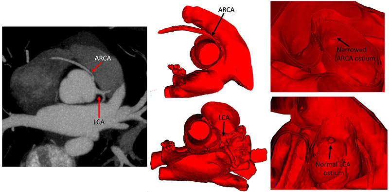
Cardiac CT image (left) demonstrates an anomalous right coronary artery (ARCA) and a normal left coronary artery (LCA). 3D digital modeling of the ARCA and LCA clarify the spatial anatomy as seen from the inside of the heart (middle). Ostial narrowing of the ARCA is clearly seen from the inside of the aorta on the 3D model (right). (Courtesty of Dr. Kanwal Farooqi)
According to the authors, the 3D models were much more accurate in demonstration of a separate AAOCA ostium, 97.4 percent vs. 65 percent on echo and 67 percent by CT. The accuracy of assessment for ostial stenosis was also highest for 3D models, albeit at 56 percent.
“Our 3D digital models, which can also be 3D printed, were able to capture the opening of the coronary artery more clearly, providing an enhanced view of the ostium,” explains Dr. Farooqi. “When this area is narrowed, we believe that the patient is at increased risk for sudden cardiac death. It is challenging to decide if surgery should be performed in patients with anomalous right coronary arteries. The subtle information we gain from 3D digital models augments other clinical data to help us make these critical decisions.”
“Our 3D digital models, which can be printed, were able to capture the opening of the coronary artery more clearly, providing an enhanced view of the ostium.” — Dr. Kanwal Farooqi
“There is a lot of controversy about whether to operate in this subset of patients with anomalous aortic origin of the right coronary artery,” says Dr. Bacha. “The imaging work of Dr. Farooqi is helping us to make this decision based on specific criteria. For example, if there is ostial narrowing, it would push us toward operating, and if the ostium does not appear stenotic, then we would not operate.”
Dr. Bacha and Dr. Farooqi are now leading efforts to establish an anomalous coronary artery program in which a core group of physicians regularly meet to discuss patient management. With a large prospective database of patients operated on and closely monitored for AAOCA in the last 10 to 15 years, physicians at NewYork-Presbyterian Morgan Stanley Children’s Hospital are able to track patients with this rare but increasingly diagnosed condition.
“We are creating a clinical program where surgeons, cardiologists, and imagers are all putting our minds together to figure out how to manage these complex patients,” says Dr. Farooqi, who is a member of the Society of Cardiovascular Computed Tomography Future Leaders Program. “We are now working on a new study examining the use of the noninvasive HeartFlow® technology in children with AAOCA. This is an innovative analysis technique for cardiac CT that can give us information, noninvasively, regarding flow within a coronary artery.”
Read More
Farooqi KM, Nees SN, Smerling J, Senapathi SH, Lorenzoni R, Pavlicova M, Einstein AJ, Bacha BA, Kalfa D, Chai PJ. Assessment of anomalous coronary arteries by imagers and surgeons: comparison of imaging modalities. The Annals of Thoracic Surgery. 2021 Feb;111(2):672-681.
NewYork-Presbyterian
Advances in Pediatrics
Read more about our latest clinical advances.