


Congenital Heart Disease Conditions
NewYork-Presbyterian provides comprehensive, lifelong care for the following types of congenital heart disease:
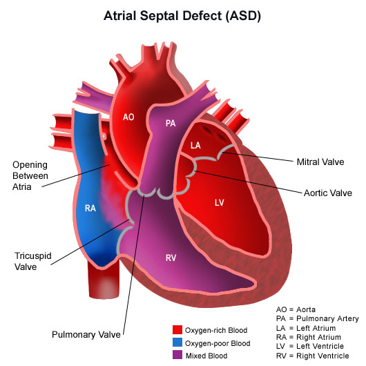
- Atrial septal defect. An opening in the wall between the right and left atria (chambers of the heart) that results in abnormal blood flow through the heart. Left untreated, atrial septal defects can cause enlargement of the right side of the heart, arrhythmia's (irregular heart rhythm), and, in some cases, pulmonary hypertension.
- Atrioventricular canals. Large openings between the right and left sides of the heart. Usually, one large common valve replaces the normal mitral valve and tricuspid valve. Left untreated, this defect can cause poor growth, malnourishment, enlargement of the heart, and pulmonary hypertension.
- Coarctation of the aorta. A constriction in the aorta that causes blood pressure to rise above the narrowed area while limiting blood flow to the body.
- Ebstein's anomaly. This disorder involves the tricuspid valve, which separates the right upper chamber (right atrium) from the right lower chamber (right ventricle) of the heart. The valve's "leaflets," which normally open to allow blood to flow from the upper to lower chamber and close to prevent it from flowing backward, do not function properly. Blood may backflow into the upper chamber and cause swelling in the heart and/or fluid buildup in the lungs or liver.
- Hypoplastic left heart syndrome. The left side of the heart is incompletely formed. This defect is typically repaired with a technique known as the "Norwood procedure." Our congenital heart surgeons are among the most experienced in the country performing this procedure, and we achieve some of the best outcomes.
- Patent ductus arteriosus. Ductus arteriosus is a vessel that allows blood to bypass a baby's lungs before birth. Within a few days after birth, the vessel typically closes. But in babies with patent ductus arteriosus, the vessel remains open and interferes with blood flow between the aorta and pulmonary artery.
- Pulmonary artery stenosis. A narrowing of the pulmonary artery, which carries blood into the lungs so that they may be infused with oxygen. Without enough oxygen-rich blood, the body cannot function properly. To overcome the lack of oxygen-rich blood, the heart tries to push more blood through the pulmonary artery, which can raise pressure in the right ventricle and damage the heart.
- Single ventricle. Single ventricle is a collective term to describe defects such as hypoplastic left heart syndrome, in which oxygen-rich and oxygen-poor blood mix in a single chamber of the heart. Our heart surgeons have great expertise performing the Fontan procedure, which directs oxygen-poor blood directly to the pulmonary artery and lungs, and provide lifelong care for people born with single ventricle disorders.
- Sinus of Valsalva aneurysm. A ruptured sinus of Valsalva aneurysm causes a communication between the aorta and the atrium or right ventricle. This abnormality is often congenital, but may also result from endocarditis (inflammation of the inner lining of the heart) or trauma. Surgery is the recommended treatment and requires special expertise, such as that found at NewYork-Presbyterian.
- Tetralogy of Fallot. The most common cyanotic defect (where the heart delivers less oxygen-rich blood to the body than normal), this complex congenital condition consists of four developmental defects which need to be surgically repaired early in childhood. Many people with this disorder also need the pulmonary valve replaced when they are adults. We are renowned for our experience treating patients with Tetralogy of Fallot.
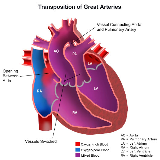
- Transposition of the great arteries. The anatomical positions of the pulmonary artery and aorta are switched, so that the aorta arises from the right ventricle and the pulmonary artery arises from the left ventricle. This causes oxygen-poor blood to be circulated to the body instead of oxygen-rich blood, a life-threatening medical emergency that requires immediate treatment. Our surgeons have pioneered the innovative "arterial switch procedure," which recreates normal anatomy and function while reducing the risk of complications associated with other surgeries.
- Valve repair and re-repair. It is not unusual for people who've had valve repair surgery for congenital heart disease as children to require another surgical repair or replacement as adults. Our surgeons have exceptionally high experience performing these procedures. Examples include repair or replacement of the aortic valve in patients with congenital aortic stenosis or other disorders affecting this valve, and repair or replacement of the pulmonary valve in people with pulmonary atresia (a form of heart disease in which the pulmonary valve does not form properly).
- Ventricular septal defect. An opening in the wall that separates the two ventricles of the heart, causing oxygen-poor blood to mix with oxygen-rich blood. Our surgeons are highly skilled in correcting these defects, using robotic surgery for some patients.